Figures & data
Figure 1 Mean ( ± SEM) plasma progesterone in gonadally intact (upper panel) and castrated (lower panel) prepubertal (28 days old) and adult (77 days old) males either before or after a 30 min restraint stress. Asterisks indicate a significant difference between prepubertal and adult males (P < 0.05).
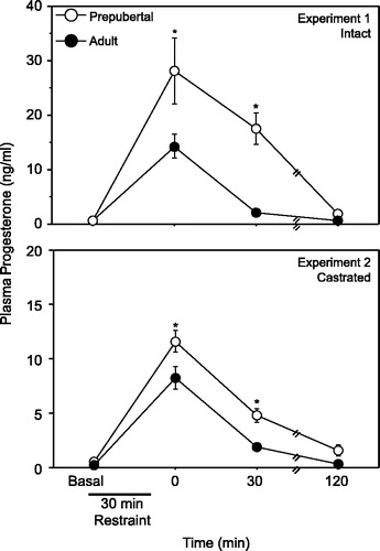
Table I. Mean (±SEM) % adrenal/body weight of prepubertal (28 days old) and adult (77 days old) males in experiments 1 and 2.
Figure 2 Mean ( ± SEM) PR-ir cell number (cells/40,000 μm2) in the PVN of gonadally intact, unstressed prepubertal and adult male rats. Asterisk indicates a significant difference between prepubertal and adult males (P < 0.05).
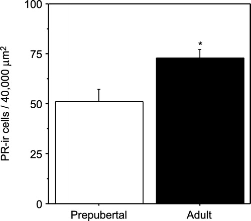
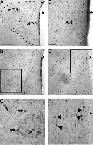
Figure 3 Photomicrographs of PR-ir cells in the PVN of prepubertal and adult males, and the AH (AH; panel D) of an adult male for comparison. (A) low power (10 × ) picture of the PVN and magnocellular and parvocellular subdivisions of the PVN in a prepubertal male. A higher power (20 × ) picture of the PVN in a prepubertal (B) or adult (E) male, while boxes are the areas of higher magnification (40 × ) in the prepubertal (C) and adult (F) male shown below. Arrows in panel C represent immunopositive cells in the prepubertal PVN, while arrowheads in panel F represent immunopositive cells in the adult PVN. (A) Bar = 150 μm, (B, D and E) Bar = 50 μm and (C and F) Bar = 25 μm. Abbreviations: AH, anterior hypothalamus; mPVN, magnocellular subdivision of the PVN; pPVN, parvocellular subdivision of the PVN; asterisks indicate the third ventricle.