Figures & data
Figure 1 (A) RGS4 mRNA expression in the hippocampal subfields (CA1, CA3 and DG) was not significantly affected by an injection with a high dose of corticosterone (CA1: P = 0.17; CA3: P = 0.39; DG: P = 0.18). (B) Autoradiogram of a complete coronal brain section from a vehicle-injected animal (left) shows the overall expression pattern of RGS4 mRNA in the rat brain. On the right, hybridization signals of the hippocampus are shown separately for a vehicle- and a corticosterone-injected animal. Hybridization with the sense probe did not yield any specific signal (data not shown). N = 8 per group.
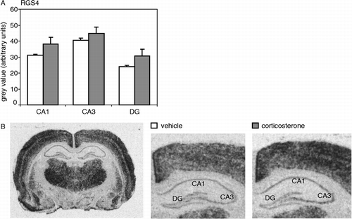
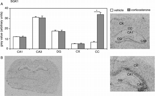
Figure 2 (A) SGK1 mRNA expression in several brain areas after a vehicle or corticosterone injection. Expression in the hippocampal subfields (CA1, CA3 and DG), or in the CX, was not affected by corticosterone (CA1: P = 0.75; CA3: P = 0.84; DG: P = 0.78; CX: P = 0.99). In the CC, SGK1 mRNA expression was significantly (P < 0.001, indicated by an asterisk) increased 1 h following a single injection with a high dose of corticosterone. (B) Autoradiogram of a complete coronal brain section from a vehicle-injected animal (left) shows the overall expression pattern of SGK1 mRNA in the rat brain. On the right, hybridization signals of the hippocampus and adjacent CC are shown separately for a vehicle- and a corticosterone-injected animal. Hybridization with the mismatch probe did not yield any specific signal (data not shown). N = 8 per group.