Figures & data
Figure 1 (a) Time course of changes in [Ca2 + ]i in response to Con A (2 μg/ml) in splenic lymphocytes from control and acute restraint-stressed mice; (b) effects of Con A and Con A plus verapamil on [Ca2 + ]i in splenic lymphocytes from control (n = 7 per group) and acute restraint-stressed (n = 9 per group) mice. Fura-2-loaded lymphocytes were incubated for 8 min with or without Con A. Verapamil was added 10 min before exposure to Con A. The net Δ[Ca2 + ]i is the difference in the increase in [Ca2 + ]i 8 min after addition of Con A or Con A plus verapamil, vs. the vehicle. Data are presented as group means ± SEM. Statistically significant differences from control are indicated as: *P < 0.05, **P < 0.01.
![Figure 1 (a) Time course of changes in [Ca2 + ]i in response to Con A (2 μg/ml) in splenic lymphocytes from control and acute restraint-stressed mice; (b) effects of Con A and Con A plus verapamil on [Ca2 + ]i in splenic lymphocytes from control (n = 7 per group) and acute restraint-stressed (n = 9 per group) mice. Fura-2-loaded lymphocytes were incubated for 8 min with or without Con A. Verapamil was added 10 min before exposure to Con A. The net Δ[Ca2 + ]i is the difference in the increase in [Ca2 + ]i 8 min after addition of Con A or Con A plus verapamil, vs. the vehicle. Data are presented as group means ± SEM. Statistically significant differences from control are indicated as: *P < 0.05, **P < 0.01.](/cms/asset/11e99651-8e54-4750-a62c-c5562dbc7367/ists_a_209523_f0001_b.gif)
Figure 2 (a) Time course of changes in [Ca2 + ]i in response to LPS (25 μg/ml) in splenic lymphocytes from control and acute restraint-stressed mice; (b) effect of LPS on [Ca2 + ]i in splenic lymphocytes from control (n = 6 per group) and acute restraint-stressed (n = 7 per group) mice. Fura-2-loaded lymphocytes were incubated for 8 min with or without LPS. The net Δ[Ca2 + ]i is the difference in the increase in [Ca2 + ]i 8 min after addition of LPS vs. vehicle. Data are presented as group means ± SEM.
![Figure 2 (a) Time course of changes in [Ca2 + ]i in response to LPS (25 μg/ml) in splenic lymphocytes from control and acute restraint-stressed mice; (b) effect of LPS on [Ca2 + ]i in splenic lymphocytes from control (n = 6 per group) and acute restraint-stressed (n = 7 per group) mice. Fura-2-loaded lymphocytes were incubated for 8 min with or without LPS. The net Δ[Ca2 + ]i is the difference in the increase in [Ca2 + ]i 8 min after addition of LPS vs. vehicle. Data are presented as group means ± SEM.](/cms/asset/4c112b5a-8f83-4e90-a11a-2760eb702917/ists_a_209523_f0002_b.gif)
Figure 3 Proliferative effects of Con A and Con A plus verapamil on splenic lymphocytes from control (n = 7 per group) and acute restraint-stressed (n = 7 per group) mice. Lymphocytes were stimulated for 42 h with Con A or Con A plus verapamil, and proliferation was measured by MTT assay. Verapamil was added 10 min before exposure to Con A. Data are presented as group means ± SEM. Statistically significant differences vs. control are indicated as: *P < 0.05, **P < 0.01. OD570, optical density at 570 nm.
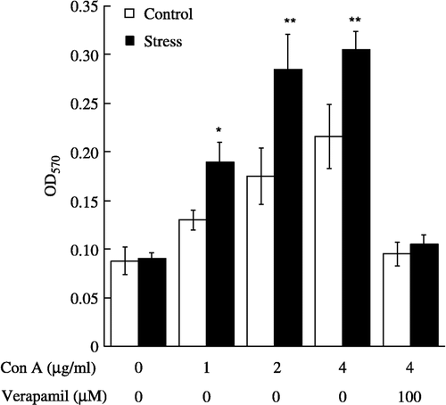
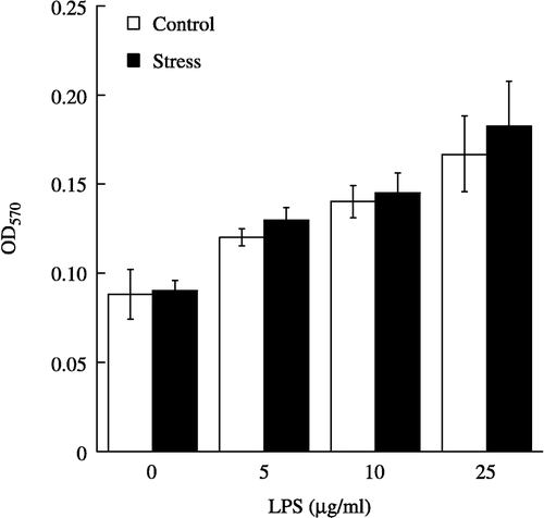
Figure 4 Proliferative effect of LPS on splenic lymphocytes from control (n = 7 per group) and acute restraint-stressed (n = 7 per group) mice. Lymphocytes were stimulated for 42 h with various concentrations of LPS, and proliferation was measured by MTT assay. Data are presented as group means ± SEM. OD570, optical density at 570 nm.