Figures & data
Figure 1 T10 spinal cord. Coronal section. Ba-Prv virus-labeled neurons in the IML cell column 3.5 day after inoculation. Abbreviations: C, central canal; df, dorsal funiculus; dh, dorsal horn; lf, lateral funiculus; vh, ventral horn. Scale bar = 200 μm.
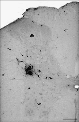
Figure 2 Virus-infected cells in the hypothalamus (A,B) 5 days, and in the pons (C,D) and the medulla oblongata (E,F) 4 days after inoculation. (A) Paraventricular nucleus, dorsolateral parvicellular part. (B) Scattered cells in the lateral hypothalamic area and the arcuate nucleus. (C) Caudal part of the locus coeruleus, subcoeruleus area and the rostral part of the A5 cell group. (D) Caudal part of the pons: A5 cell group. (E) Labeled cells in the RVMM and scattered labeled cells in the C3 adrenaline cell group. (F) RVLM. Labeled cells are concentrated in the rostral part of the C1 adrenaline cell group. Abbreviations: Ar, arcuate nucleus; A5, A5 noradrenaline cell group; Ce, central amygdaloid nucleus; IO, inferior olive; LH, lateral hypothalamic area; p, pyramidal tract; PGi, paragigantocellular reticular nucleus; PVN, paraventricular nucleus; SC, subcoeruleus area; tp, trapezoid body; 3V, third ventricle; 4V, 4th ventricle; 7th, cranial portion of the facial nerve. Scale bars = 500 μm (A) μm and 1 mm (B–F).
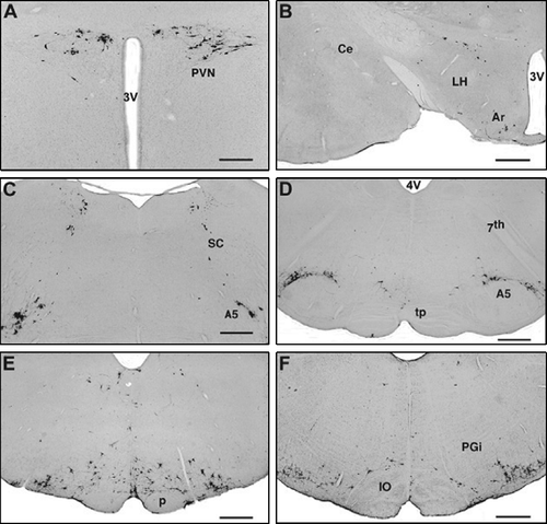
Table I. Number of Ba-Prv virus-infected cells (4 and 5 days after inoculation) in brain regions in intact (right and left side) and neurosurgically-operated rats (ipsi- and contra-lateral sides to the unilateral subcutaneous injection).
Figure 3 Unilateral (right side) spino-medullary hemisection, ipsilateral (right side) viral injections into the hindlimb (A–C). Four/five days after inoculation, infected cells appear only on the contralateral (left) side in the hypothalamic paraventricular nucleus (A), the pons (B) and the rostral medulla oblongata (C). Unilateral (left side) spino-medullary hemisections, contralateral to viral injection into the right hind limb (D–F). Four/five days after inoculation, infected cells appear only on the side of the injection (right) in the hypothalamic paraventricular nucleus (D), the pons (E) and the rostral medulla oblongata (F). Abbreviations: A5, A5 noradrenaline cell group; Ce, central amygdaloid nucleus; Gi, gigantocellular reticular nucleus; IO, inferior olive; LC, locus coeruleus; ot, optic tract; PVN, paraventricular nucleus; SC, subcoeruleus area; 3V, third ventricle. Scale bars = 500 μm (A) and 1 mm (B–F).

Figure 4 Midsagittal medullary transections. Bilateral distribution of viral infected cells (similar to unoperated rats) in the hypothalamic paraventricular nucleus (A) 5 days and (B) in the rostral medulla oblongata (arrows point to the dense populations of infected cells in the C1 cell group) 4 days after inoculation of the virus into the right hind limb. Abbreviations: p, pyramidal tract; PGi, paragigantocellular reticular nucleus; PVN, paraventricular nucleus; 3V, third ventricle. The arrowheads point to the mid-sagittal medullary transection (narrow vertical cut in the midline). Scale bars = 500 μm (A) and 1 mm (B).
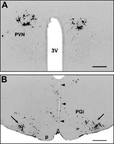
Figure 5 Descending supraspinal pathways to the sympathetic preganglionic neurons in the spinal cord and their possible cross over in the spinal cord. Fibers arise in the hypothalamus (neurons in the paraventricular [PVN] and arcuate nuclei, and the lateral hypothalamic area [LH]), the pons (neurons in the A5, A7 cell groups, caudal portion of the locus coeruleus [LC] and the subcoeruleus area) and in the medulla oblongata (neurons in the C1 adrenaline cell group, the RVMM and the nucleus of the solitary tract). After cross over in the spinal cord, fibers terminate on the sympathetic preganglionic neurons in the IML cell column of the T10 spinal cord. 1, site of the Ba-Prv viral injection (primary nociceptive neurons); 2, unilateral transection between the spinal cord and the medulla oblongata; 3, midsagittal transection through the medulla oblongata.
![Figure 5 Descending supraspinal pathways to the sympathetic preganglionic neurons in the spinal cord and their possible cross over in the spinal cord. Fibers arise in the hypothalamus (neurons in the paraventricular [PVN] and arcuate nuclei, and the lateral hypothalamic area [LH]), the pons (neurons in the A5, A7 cell groups, caudal portion of the locus coeruleus [LC] and the subcoeruleus area) and in the medulla oblongata (neurons in the C1 adrenaline cell group, the RVMM and the nucleus of the solitary tract). After cross over in the spinal cord, fibers terminate on the sympathetic preganglionic neurons in the IML cell column of the T10 spinal cord. 1, site of the Ba-Prv viral injection (primary nociceptive neurons); 2, unilateral transection between the spinal cord and the medulla oblongata; 3, midsagittal transection through the medulla oblongata.](/cms/asset/d7f592a0-cd47-4148-a2cf-c11a4f4eb2d8/ists_a_242355_f0005_b.gif)