Figures & data
Table I. Effects of exposure to HU on catecholamine levels measured in plasma and spleen.
Figure 1 Effects of exposure to HU conditions on spleen size. Mice were normally caged (NC, white bar) or exposed to HU for 18 h (HU-18, grey stippled bar), or 48 h (HU-48, black bar). Spleen size is expressed as the means of spleen weights (gram) divided by body weights (gram) for each mouse ± SE. * indicates significant difference compared to normally caged group (*p ≤ 0.05, **p ≤ 0.01). # indicates significant difference between hindlimb unloaded time point groups (p ≤ 0.05).
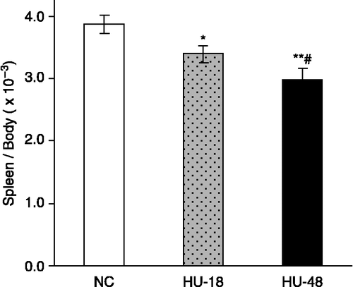
Figure 2 Effects of exposure to HU on numbers of TEPC. Mice were injected with 2 ml of sterile 3% thioglycollate, an inflammatory agent, into the peritoneal cavity four days prior to harvest and normally-caged (NC, white bar) or exposed to HU for 18 h (HU-18, grey stippled bar) or 48 h (HU-48, black bar) prior to harvest. Values are expressed as the means of total numbers of peritoneal cells collected ± SE. Asterisk indicates significant difference compared to normally caged group (p ≤ 0.05).
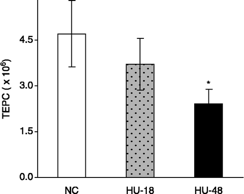
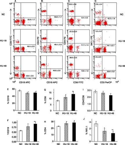
Figure 3 Effects of exposure to HU conditions on splenic immune cell type percentage distribution. Samples of 106 resuspended spleen cells from normally caged mice (NC, white bar) or mice HU-exposed for 18 h (HU-18, grey stippled bar), or 48 h (HU-48, black bar) were stained with CD8α-FITC, CD4-PE, CD19-APC (A–C); CD8α-FITC, NK1.1-PE, CD3-PerCP (D); or matched isotype controls. (A–D) Flow cytometric dot plots; inset represents mean percent ± SE. (E–J) Bar graphs; values are recorded as percent ± SE of cells within the lymphocyte gate stained positive for the indicated surface antigen. In (F), “T” is defined as percent CD4+ plus CD8+ cells. Asterisks indicate significant difference compared to normally caged group (*p ≤ 0.05, **p ≤ 0.01).