Figures & data
Table I. Behavioral alterations induced by chronic restraint stress.
Figure 1 Effects of chronic stress on lymphoproliferative response to selective T- and B- mitogens. Cells from lymph nodes and spleens of stressed mice and untreated controls were stimulated with Con A and LPS respectively, labeled with [3H]thymidine for 72 h and harvested. Representative results from three independent experiments are shown. Values are expressed as group means ± SEM. of the stimulated/basal ratio (proliferation index). Statistical significance was determined using unpaired t-test (n = 6 mice per group, **p < 0.01).
![Figure 1 Effects of chronic stress on lymphoproliferative response to selective T- and B- mitogens. Cells from lymph nodes and spleens of stressed mice and untreated controls were stimulated with Con A and LPS respectively, labeled with [3H]thymidine for 72 h and harvested. Representative results from three independent experiments are shown. Values are expressed as group means ± SEM. of the stimulated/basal ratio (proliferation index). Statistical significance was determined using unpaired t-test (n = 6 mice per group, **p < 0.01).](/cms/asset/de9c8118-c09e-4726-bcf7-9d8fdf58507f/ists_a_313909_f0001_b.gif)
Figure 2 Effects of chronic stress on NK activity. Cells suspensions from spleens of stressed mice and untreated controls were co-incubated with YAC-1 cells labeled with [3H]-thymidine at different target:effector ratios, cultured for 3.5 h and harvested. Representative results from three independent experiments are shown. Values are expressed as group means ± SEM. of NK specific cytolysis, calculated as 100 × (SR-ER)/SR, where SR is the spontaneous release and ER is the experimental release of [3H]thymidine. Statistical significance was determined using unpaired t-test (n = 4 mice per group, no significant difference).
![Figure 2 Effects of chronic stress on NK activity. Cells suspensions from spleens of stressed mice and untreated controls were co-incubated with YAC-1 cells labeled with [3H]-thymidine at different target:effector ratios, cultured for 3.5 h and harvested. Representative results from three independent experiments are shown. Values are expressed as group means ± SEM. of NK specific cytolysis, calculated as 100 × (SR-ER)/SR, where SR is the spontaneous release and ER is the experimental release of [3H]thymidine. Statistical significance was determined using unpaired t-test (n = 4 mice per group, no significant difference).](/cms/asset/66bdae8c-f959-4a20-8b66-e21a8ca8cb63/ists_a_313909_f0002_b.gif)
Table II. T lymphocyte subsets distribution in stressed and control mice.
Figure 3 Effects of chronic stress on TNF-α and IFN-γ mRNA expression in lymphocytes. Total RNA and mRNA were isolated from lymph nodes of stressed mice, and untreated mice, and used for cDNA synthesis. Cytokine expression was evaluated by Real Time RT-PCR using SYBR Green dye. Representative results from three independent experiments are shown. Values are expressed as group means ± SEM. normalized with β-actin as housekeeping gene. Statistical significance was determined using unpaired t-test (n = 5 mice per group, **p < 0.01).
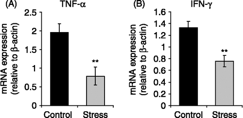
Figure 4 Effect of chronic stress on LBC lymphoma cell proliferation and prognosis. Mice were subjected to chronic restraint stress, or control conditions, for three weeks, and then were inoculated subcutaneously with 1 × 10⁶ LBC cells to generate a solid tumor. Stress procedures continued until the mice died. Representative results from three independent experiments are shown. (A) Tumor progression after appearance. Tumor length and width were measured and tumor volume was calculated as V = π/6 × L × W2. Values are expressed as group means ± SEM. for each day post-tumor injection (p.t.i.). Statistical significance was determined using unpaired t-test (n = 10 mice per group, **p < 0.01). (B) CFSE fluorescence intensity of LBC cells growing in stressed and control mice. The graph shows the number of events against the mean fluorescence intensity for each treatment, for a representative experiment of one stressed and one control mouse. Statistical significance was determined using unpaired t-test (n = 3 mice per group, p < 0.01). (C) Kaplan-Meier plot of survival of stressed and control mice (n = 10 mice per group, p < 0.01, Log-Rank analysis).
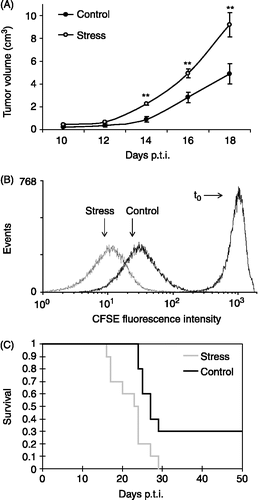
Figure 5 Effects of chronic stress on specific cytotoxic activity against LBC cells. Cells from spleens of stressed mice and untreated controls were co-cultured with LBC cells labeled with [3H]thymidine, and its release was evaluated as an indicator of cytolysis. Representative results from three independent experiments are shown. Values are expressed as group means ± SEM. of the percentage of cytotoxic activity at different effector:target ratios. Statistical significance was determined using unpaired t-test (n = 4 mice per group, *p < 0.05, **p < 0.01).
![Figure 5 Effects of chronic stress on specific cytotoxic activity against LBC cells. Cells from spleens of stressed mice and untreated controls were co-cultured with LBC cells labeled with [3H]thymidine, and its release was evaluated as an indicator of cytolysis. Representative results from three independent experiments are shown. Values are expressed as group means ± SEM. of the percentage of cytotoxic activity at different effector:target ratios. Statistical significance was determined using unpaired t-test (n = 4 mice per group, *p < 0.05, **p < 0.01).](/cms/asset/6d943518-2f94-4026-8d4d-0b407eacbe8e/ists_a_313909_f0005_b.gif)