Open access
466
Views
0
CrossRef citations to date
0
Altmetric
Abstract
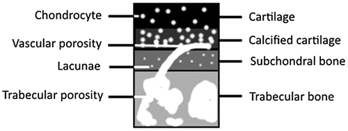
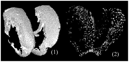
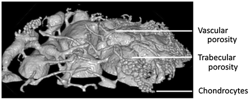
3D morphological analyses of subchondral mineralized zone in mice knee joint from HR-μCT scan
B. MehadjiDepartment of Physical Therapy, College of Staten Island, City University of New York, New York, NY, USACorrespondence[email protected]
& J. P. BerteauDepartment of Physical Therapy, College of Staten Island, City University of New York, New York, NY, USA
Pages S129-S130
|
Published online: 27 Oct 2017
Related research
People also read lists articles that other readers of this article have read.
Recommended articles lists articles that we recommend and is powered by our AI driven recommendation engine.
Cited by lists all citing articles based on Crossref citations.
Articles with the Crossref icon will open in a new tab.


