Figures & data
Figure 1. Generating 3D model of inner ear (A, B) the mask of the inner ear in Mimics 20.0; (C, D) the reconstruction model of 3D structure of inner ear; (E, F) the finite element mesh.
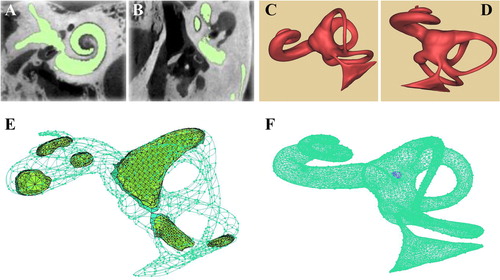
Figure 2. Parameters on the 3D model (A, B) the narrowest cross section of the inner ear model; (C) the time step-pressure function for the simulated external forces; (D) the speed-displacement curve under different grids.
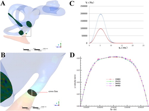
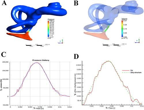
Figure 3. The simulated biomechanical properties of the 3D model (A) pressure cloud of the bony labyrinth in the inner ear; (B) velosity cloud of the bony labyrinth in the inner ear; (C) pressure variation curve of inlet surface and fluid-solid coupled surface; (D) displacement curve of the round membrane.