Figures & data
Figure 1. Installation of works for the matter of flux exhibition at art laboratory Berlin, including objects from the mooncalf project. © WhiteFeather Hunter, Citation2023.
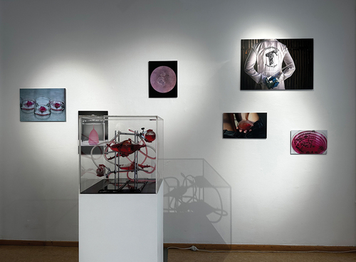
Figure 2. Video still of biohazardous materials transport by bicycle. © WhiteFeather Hunter, Citation2023.
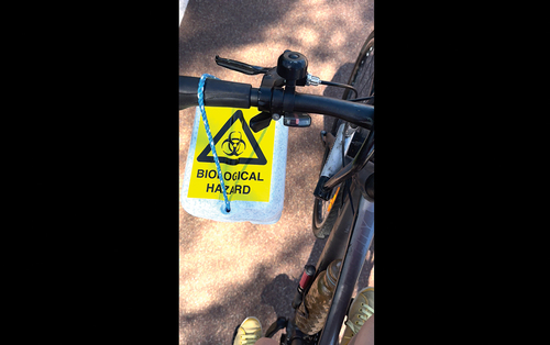
Figure 3. Endometrial tissue explant. © WhiteFeather Hunter, Citation2022.
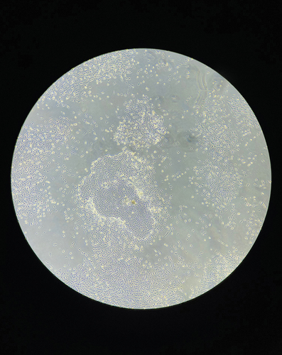
Figure 4. Separated layers of menstrual fluid. © WhiteFeather Hunter, Citation2022.
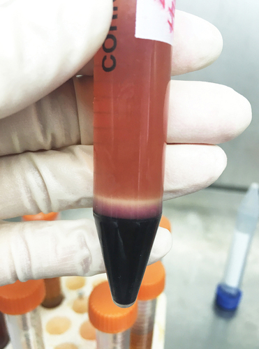
Figure 5. Happiness is a Warm Gun, machine embroidered lab coat stained with menstrual fluid. © WhiteFeather Hunter, Citation2022.
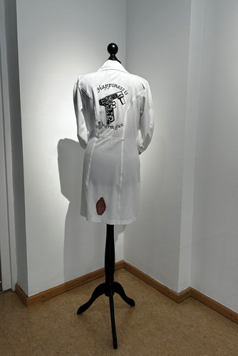
Figure 6. Tissue culture flasks with menstrual-derived cells. © WhiteFeather Hunter, Citation2023.
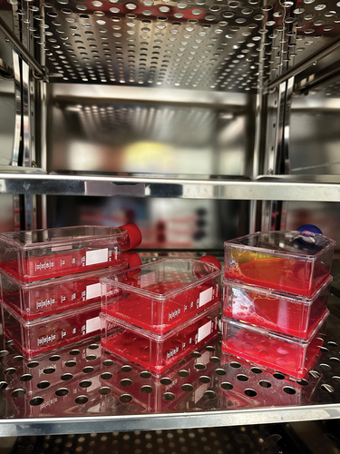
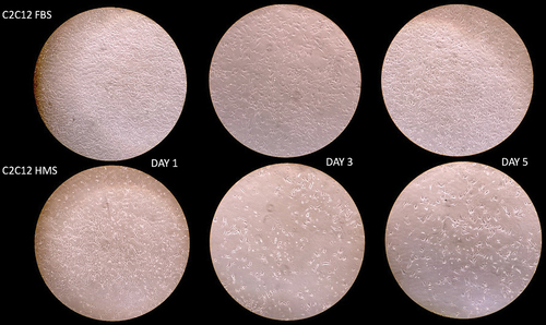
Figure 7. Comparative microscopy showing reduced cell adhesion over a five-day period, in C2C12 cells grown in human menstrual serum (HMS) versus those grown in fetal bovine serum (FBS) © WhiteFeather Hunter, Citation2022.