6,262
Views
0
CrossRef citations to date
0
Altmetric
Invited Reviews

Free thyroxine measurement in clinical practice: how to optimize indications, analytical procedures, and interpretation criteria while waiting for global standardization

ORCID Icon, , ORCID Icon, ORCID Icon & ORCID Icon
Pages 101-140 | Received 28 Mar 2022, Accepted 03 Sep 2022, Published online: 13 Oct 2022

Figures & data

Figure 1. Chemical structure of thyroid hormones.

Figure 1. Chemical structure of thyroid hormones.

Figure 2. Biosynthesis of thyroid hormones. (T4, thyroxine, T3, triiodothyronine, MCT8, monocarboxylate transporter 8; Tg, thyroglobulin; TPO, thyroid peroxidase; DIT, diiodotyrosine; MIT, monoiodotyrosine).

Figure 2. Biosynthesis of thyroid hormones. (T4, thyroxine, T3, triiodothyronine, MCT8, monocarboxylate transporter 8; Tg, thyroglobulin; TPO, thyroid peroxidase; DIT, diiodotyrosine; MIT, monoiodotyrosine).

Figure 3. The log-linear inverse relationship between TSH and FT4. (TSH, thyroid stimulating hormone; FT4, free thyroxine). The blue line represents an approximate relationship between TSH and FT4. The dotted grey lines represent the normal values for TSH (horizontal) and FT4 (vertical).

Figure 3. The log-linear inverse relationship between TSH and FT4. (TSH, thyroid stimulating hormone; FT4, free thyroxine). The blue line represents an approximate relationship between TSH and FT4. The dotted grey lines represent the normal values for TSH (horizontal) and FT4 (vertical).

Table 1. Clinical indications for FT4 measurement.

Figure 4. Schematic of the four main immunoassay formats used to measure FT4. (A) In the two-step labeled T4 analog method, FT4 in the specimen interacts with anti-hormone antibodies on a solid support. Antibody binding sites are occupied by endogenous FT4. The specimen is then removed and any empty antibody binding sites are allowed to interact with labeled FT4 (“back-titration”). (B) In the one-step assay, FT4 in the specimen competes with the labeled analog for binding to the antibodies fixed on a solid phase. The amount of labeled hormone left after the final washing of the solid support is inversely proportional to the original concentration of free hormone in the sample. (C) In the one-step analog two-step incubation format, anti-T4 antibodies are first incubated with FT4 in the specimen. In the second incubation step, the labeled T4 analog and streptavidin-coated microparticles bind to the unoccupied anti-T4 antibody binding sites, forming an antibody-hapten complex, bound to the solid phase via interaction between biotin and streptavidin. The unbound labeled T4 analog is washed away and the signal recorded. (D) In the labeled antibody method, labeled T4 antibodies bind to either FT4 in the specimen or solid phase-bound T4. After a short incubation period, a wash is carried out and the signal is measured (FT4, free thyroxine; T4, thyroxine).

Figure 4. Schematic of the four main immunoassay formats used to measure FT4. (A) In the two-step labeled T4 analog method, FT4 in the specimen interacts with anti-hormone antibodies on a solid support. Antibody binding sites are occupied by endogenous FT4. The specimen is then removed and any empty antibody binding sites are allowed to interact with labeled FT4 (“back-titration”). (B) In the one-step assay, FT4 in the specimen competes with the labeled analog for binding to the antibodies fixed on a solid phase. The amount of labeled hormone left after the final washing of the solid support is inversely proportional to the original concentration of free hormone in the sample. (C) In the one-step analog two-step incubation format, anti-T4 antibodies are first incubated with FT4 in the specimen. In the second incubation step, the labeled T4 analog and streptavidin-coated microparticles bind to the unoccupied anti-T4 antibody binding sites, forming an antibody-hapten complex, bound to the solid phase via interaction between biotin and streptavidin. The unbound labeled T4 analog is washed away and the signal recorded. (D) In the labeled antibody method, labeled T4 antibodies bind to either FT4 in the specimen or solid phase-bound T4. After a short incubation period, a wash is carried out and the signal is measured (FT4, free thyroxine; T4, thyroxine).

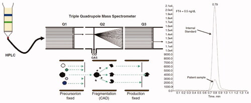
Table 2. Main analytical characteristics of the most used FT4 immunoassays as quoted by the manufacturers.

Figure 5. Schematic depiction of the workflow for current (high-pressure) liquid chromatography-tandem mass spectrometry (LC-MS/MS) of FT4.

Figure 5. Schematic depiction of the workflow for current (high-pressure) liquid chromatography-tandem mass spectrometry (LC-MS/MS) of FT4.

Table 3. Comparison of RIs of FT4 in adult populations on Abbott ARCHITECT platform.

Table 4. Comparison of RIs of FT4 in adult populations on Beckman Coulter platforms.

Table 5. Comparison of reference intervals of FT4 in adult populations on Roche Diagnostics platforms.

Table 6. Comparison of reference intervals of FT4 in adult populations on the Siemens ADVIA Centaur platform.

Table 7. Comparison of RIs of FT4 in elderly populations using different analytical platforms.

Figure 6. FT4 concentrations during pregnancy and post-partum. Error bars represent the 5th and 95th percentiles per trimester. Dashed lines represent non-pregnant lower and upper reference limits. (Green, 1st tertile; violet, 2nd tertile; grey, 3rd tertile; *p < 0.0001 compared with Tr1; **p < 0.01 compared with Tr1; NS, not statistically significant compared with Tr1m; FT4, free thyroxine; NS, non-significant; Tr1, trimester 1; Tr2, trimester 2; Tr3, trimester 3; TSH, thyroid-stimulating hormone; PP, post-partum).

Figure 6. FT4 concentrations during pregnancy and post-partum. Error bars represent the 5th and 95th percentiles per trimester. Dashed lines represent non-pregnant lower and upper reference limits. (Green, 1st tertile; violet, 2nd tertile; grey, 3rd tertile; *p < 0.0001 compared with Tr1; **p < 0.01 compared with Tr1; NS, not statistically significant compared with Tr1m; FT4, free thyroxine; NS, non-significant; Tr1, trimester 1; Tr2, trimester 2; Tr3, trimester 3; TSH, thyroid-stimulating hormone; PP, post-partum).

Table 8. Comparison of gestational RIs of FT4 using different platforms, in studies published from January 2012 to May 2022 including at least two different trimesters of pregnancy.

Figure 7. Age-dependent scatter plot of FT4 concentration, stratified by sex. (FT4, free thyroxine).

Figure 7. Age-dependent scatter plot of FT4 concentration, stratified by sex. (FT4, free thyroxine).

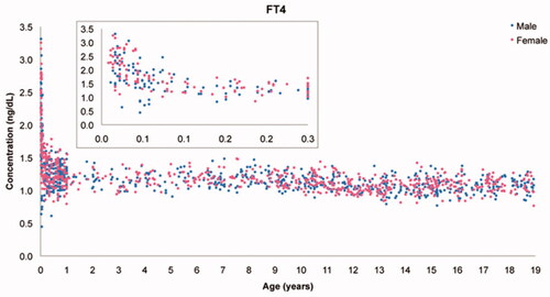
Table 9. Comparison of RIs of FT4 for children aged 1–18 years using different platforms, in studies published from January 2012 to May 2022.

Table 10. Comparison of RIs of FT4 for infants aged 0–12 months using different platforms, in studies published from January 2012 to May 2022.