Figures & data
Figure 1. Contemporary pathophysiological model of NAFLD. Environmental and genetic factors challenge homeostasis and lead to several pathological processes: metabolic disorders, oxidative stress, and inflammation. These processes also fortify each other and cause a disbalance, leading to the development of steatosis and NASH. NASH can eventually progress to fibrosis and cirrhosis.
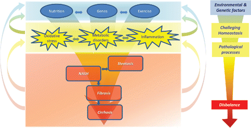
Figure 2. “Traditional” concept of action of drugs versus the contemporary concept of action of bioactives such as flavonoids. While traditional drugs are developed to act on one target, leading to absence of disease, flavonoids act on multiple targets, affecting diverse pathological processes, leading to increased ability to adapt. This fits seamlessly in the pathophysiologic model of NAFLD, since diverse pathological processes are involved ().
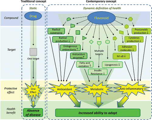
Figure 3. Schematic view of the pathophysiological pathways involved in NAFLD. Overview of the processes in the liver, adipose, and other tissues that are responsible for metabolic disturbances, oxidative stress, and inflammation in NAFLD. FFA = free fatty acids, SREBP-1c = sterol regulatory element-binding protein 1c, ChREBP = carbohydrate-responsive element-binding protein, FAS = fatty acid synthase, ACC = acetyl-CoA carboxylase, gk = glucokinase, VLDL = very low-density lipoprotein, ER = endoplasmic reticulum.
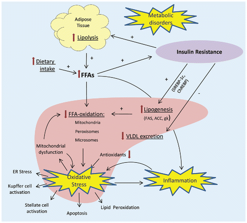
Figure 4. Major subclasses of flavonoids found in significant amounts in the pictured fruits and vegetables.
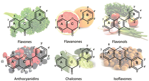
Figure 5. Effects of PPARγ stimulation by flavonoids. PPARγ stimulation by flavonoids (F) leads to changes in the factors secreted by adipose tissue, stimulating insulin sensitivity. Furthermore, it leads to the upregulation of specific genes, stimulating lipid storage in subcutaneous adipose tissue instead of the liver. TNF-α = tumor necrosis factor α, Il-6 = interleukin 6, PEPCK = phospoenolpyruvate carboxykinase, FATP = fatty acid transport protein, IRS2 = insulin receptor substrate 2, FABP = fatty acid-binding protein, ACS = acetyl-CoA synthase, DGAT = diglyceride acyltransferase, LPL = lipoprotein lipase.
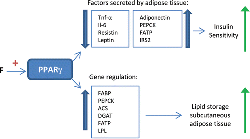
Figure 6. Effects of flavonoids on the NFκB pathway. Inhibitory effects (—|) of flavonoids (F) on different processes in the canonical NF-κB pathway, leading to a decreased inflammatory response. IKK = IκB kinase, IκBα = NF-κB inhibitor-α, p65 = NF-κB complex subunit p65, p50 = NF-κB complex subunit p50.
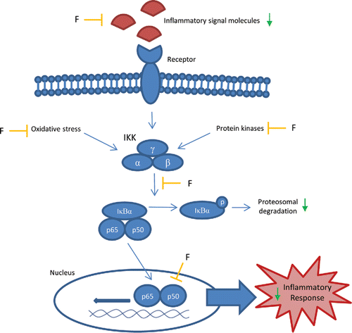
Table 1. Effects of silybin/silymarin on the development of NAFLD in animal models
Table 2. Effects of Realsil in a randomized controlled clinical trial.
Table 3. Effects of soy isoflavones on the development of NAFLD in animal models.
Table 4. Effects of green tea extract/epigallocatechin-3-gallate on the development of NAFLD in animal models.
Table 5. Effects of quercetin on the development of NAFLD in animal models.
Figure 7. Most studied flavonoids in the treatment of NAFLD. Molecular structures of the most investigated flavonoids in the treatment of NAFLD: Silybin (mixture of two diateromers, of which one is pictured), genistein, daidzein, epigallocatechin-3-gallate (ECGC), quercetin, and rutin.
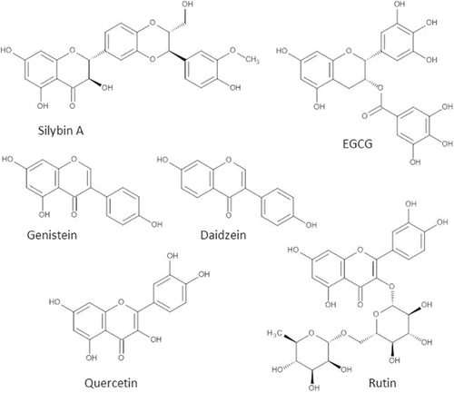
Table 6. Effects of rutin on the development of NAFLD in animal models.
Table 7. Other flavonoids investigated in animal models of NAFLD.
