7,987
Views
9
CrossRef citations to date
0
Altmetric
Review Articles

Colonization of Helicobacter pylori in the oral cavity – an endless controversy?

, , , , , , , , , & ORCID Icon show all
Pages 612-629 | Received 07 Feb 2021, Accepted 16 Mar 2021, Published online: 24 Apr 2021

Figures & data

Figure 1. Morphology of H. pylori. Field-emission scanning electron microscopic (FESEM) image of a spiral-shaped H. pylori with five to seven sheathed polar flagella. The scale bar is 0.5 μm. This figure is reprinted from references (O’Rourke Citation2001; Reshetnyak and Reshetnyak Citation2017) with kind permission from the publishers.

Figure 1. Morphology of H. pylori. Field-emission scanning electron microscopic (FESEM) image of a spiral-shaped H. pylori with five to seven sheathed polar flagella. The scale bar is 0.5 μm. This figure is reprinted from references (O’Rourke Citation2001; Reshetnyak and Reshetnyak Citation2017) with kind permission from the publishers.

Table 1. Studies published since 2010 detecting oral H. pylori with molecular methods.

Table 2. Studies published since 2010 detecting oral H. pylori with immunological and biochemical methods.

Table 3. Studies published since 2000 detecting oral H. pylori by culture techniques.
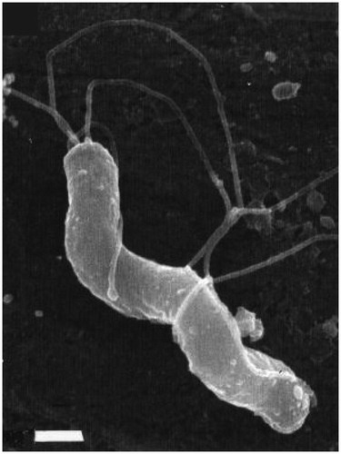
Figure 2. Morphological transformation of H. pylori from spiral to coccoid form under stress conditions. Transmission electron microscopic (TEM) images exhibiting the morphology transformation of H. pylori from its spiral to its coccoid form under stress conditions. H. pylori was cultured on Brucella blood agar with 5% horse serum and antimicrobial agents (10 mg/L vancomycin, 5 mg/L colistin, 5 mg/L trimethoprim and 5 mg/L amphotericin B) for a total of 15 days. On day 2, day 7, day 9, and day 15, its morphology was pictured by TEM: A: Day 2 (2D): bacillary form of H. pylori; B: Day 7 (7D): U-shaped form; C: Day 9 (9D): doughnut shaped form; D: Day 15 (15D): full coccoid form. The scale bar shows 1 μm. This figure is reprinted from reference (Roe et al. Citation1999) with kind permission from the publisher.

Figure 2. Morphological transformation of H. pylori from spiral to coccoid form under stress conditions. Transmission electron microscopic (TEM) images exhibiting the morphology transformation of H. pylori from its spiral to its coccoid form under stress conditions. H. pylori was cultured on Brucella blood agar with 5% horse serum and antimicrobial agents (10 mg/L vancomycin, 5 mg/L colistin, 5 mg/L trimethoprim and 5 mg/L amphotericin B) for a total of 15 days. On day 2, day 7, day 9, and day 15, its morphology was pictured by TEM: A: Day 2 (2D): bacillary form of H. pylori; B: Day 7 (7D): U-shaped form; C: Day 9 (9D): doughnut shaped form; D: Day 15 (15D): full coccoid form. The scale bar shows 1 μm. This figure is reprinted from reference (Roe et al. Citation1999) with kind permission from the publisher.

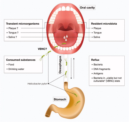
Figure 3. The cycle of H. pylori in the human body. The question on whether H. pylori is resident or transient in the oral cavity (saliva, dental plaque, or tongue microbiota) remains to be clarified. H. pylori can be introduced in the oral cavity by contaminated substances including food and drinking water. There may also be a relationship between gastric and oral H. pylori infection. H. pylori can be swallowed and may subsequently cause infections in the stomach. Conversely, reflux can bring back viable organisms, DNA fragments or antigens of H. pylori to the oral cavity from the stomach of H. pylori-infected individuals. This may lead to positive results in detection with molecular methods, immunological methods or culture methods. Furthermore, H. pylori may be present in a viable, but not-culturable (VBNC) state (i.e. coccoid form).

Figure 3. The cycle of H. pylori in the human body. The question on whether H. pylori is resident or transient in the oral cavity (saliva, dental plaque, or tongue microbiota) remains to be clarified. H. pylori can be introduced in the oral cavity by contaminated substances including food and drinking water. There may also be a relationship between gastric and oral H. pylori infection. H. pylori can be swallowed and may subsequently cause infections in the stomach. Conversely, reflux can bring back viable organisms, DNA fragments or antigens of H. pylori to the oral cavity from the stomach of H. pylori-infected individuals. This may lead to positive results in detection with molecular methods, immunological methods or culture methods. Furthermore, H. pylori may be present in a viable, but not-culturable (VBNC) state (i.e. coccoid form).

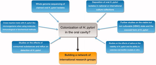
Figure 4. Future research directions. Important aspects for future studies investigating the potential colonization of H. pylori in the oral cavity: • Whole genome sequencing of oral H. pylori isolates. • Deposition of oral H. pylori in national or international culture collections. • Research on the VBNC state and the coccoid form of H. pylori. • Studies on the effects of saliva on viability of H. pylori and on the ability of H. pylori to colonize biofilms formed from oral bacteria in vitro. • Effects of consumed substances and reflux on the detection of oral H. pylori. • Cross-reaction tests with H. pylori-like microorganisms when using molecular or immunological methods. To achieve these goals, we advocate establishing a network of researchers who have reported isolating H. pylori from the oral cavity.

Figure 4. Future research directions. Important aspects for future studies investigating the potential colonization of H. pylori in the oral cavity: • Whole genome sequencing of oral H. pylori isolates. • Deposition of oral H. pylori in national or international culture collections. • Research on the VBNC state and the coccoid form of H. pylori. • Studies on the effects of saliva on viability of H. pylori and on the ability of H. pylori to colonize biofilms formed from oral bacteria in vitro. • Effects of consumed substances and reflux on the detection of oral H. pylori. • Cross-reaction tests with H. pylori-like microorganisms when using molecular or immunological methods. To achieve these goals, we advocate establishing a network of researchers who have reported isolating H. pylori from the oral cavity.