11,591
Views
6
CrossRef citations to date
0
Altmetric
Review Articles

Bdellovibrio bacteriovorus: a potential ‘living antibiotic’ to control bacterial pathogens

ORCID Icon, , ORCID Icon, ORCID Icon & ORCID Icon
Pages 630-646 | Received 20 Oct 2020, Accepted 23 Mar 2021, Published online: 01 May 2021

Figures & data

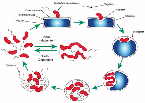
Figure 1. Schematic representation of the life cycle of B. bacteriovorus. Starting clockwise from top left of the image, the predator approaches and binds to the outer surface of its prey. The flagellum is lost and a pore is created. The predator penetrates and settles in the periplasmic space of its host. Subsequently, the pore is sealed and the predator starts consuming the intracellular components of its prey. A septation step follows, culminating in lysis of the host cell and the release of fresh B. bacteriovorus progeny. The new-born predators then start a new predation cycle either through the Host-Dependent cycle or can revert in the Host-Independent state until a suitable prey is encountered.

Figure 1. Schematic representation of the life cycle of B. bacteriovorus. Starting clockwise from top left of the image, the predator approaches and binds to the outer surface of its prey. The flagellum is lost and a pore is created. The predator penetrates and settles in the periplasmic space of its host. Subsequently, the pore is sealed and the predator starts consuming the intracellular components of its prey. A septation step follows, culminating in lysis of the host cell and the release of fresh B. bacteriovorus progeny. The new-born predators then start a new predation cycle either through the Host-Dependent cycle or can revert in the Host-Independent state until a suitable prey is encountered.

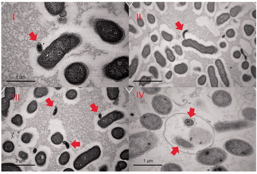
Figure 2. TEM images of various stages of predation. The prey in the images is an Enterobacter roggenkampii isolate. Images I, II and III show B. bacteriovorus HD100 (indicated with arrows) attached to the outer surface of a prey cell or in its immediate surroundings. Image IV shows a late stage of predation where the new-born predators are in the bdelloplast, prior to its disruption (our unpublished data).

Figure 2. TEM images of various stages of predation. The prey in the images is an Enterobacter roggenkampii isolate. Images I, II and III show B. bacteriovorus HD100 (indicated with arrows) attached to the outer surface of a prey cell or in its immediate surroundings. Image IV shows a late stage of predation where the new-born predators are in the bdelloplast, prior to its disruption (our unpublished data).

Table 1. Overview of bacteria known to display predatory lifestyles.

Table 2. Overview of in vivo studies that used B. bacteriovorus as a biocontrol agent against pathogens.