Figures & data
Figure 1. Time course of statin dependent effects on proliferation and AML cell viability. The time dependence of lovastatin (LST) induced effects on proliferation and apoptosis were determined in U937 or HL60 cells. Treated samples were compared with that of untreated cells. (N ≥ 3, one example is shown for each assay.) (A) Cells counts by day of culture, at various concentrations. Cell number was determined using a Coulter counter. Shown are total cell counts. At lower concentrations (i.e. <15 µM), differences between treated and untreated cells are best seen after at least 4 days of statin exposure. (B) Time course of changes in absorbance (MTS assay). Cell proliferation was measured using an MTS-like colorimetric assay that reflects cellular metabolism. (C) Effects of statins during the first 2 days of culture. Cells were plated at a concentration of 10,000 cells per well in order to look at the effects of lovastatin on proliferation during the first 2 days of drug exposure. These results show that lovastatin did not markedly affect proliferation during the first 2 days of statin exposure, especially at the lower concentrations studied. (D) Percentage of viable cells, by apoptosis, after lovastatin exposure, by day of culture (HL60 cells). Percentage of cells, by day, that were negative for both PI and Annexin. (E) Viable cell number. Using the same data set as shown in 1D, the number of viable cells was determined for each day of treatment by multiplying the number of cells as determined the Coulter counter by the percentage of cells negative for both annexin and PI. In each assay, the cytotoxic effects of concentrations of statins in the clinically relevant range (i.e. ≤5 – 12 µM) are seen only with longer duration of statin exposure.
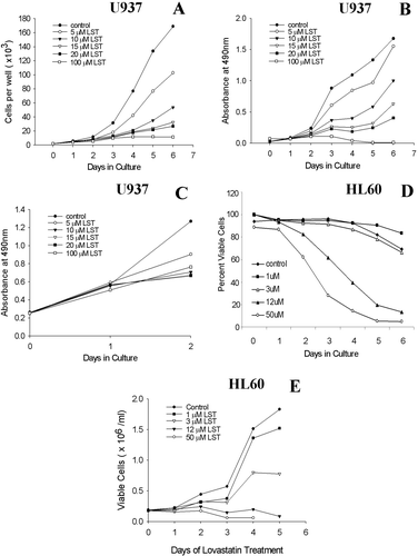
Figure 2. Lovastatin sensitivity, as measured by IC50 and IC90 determinations at days 2 and 6. The values represent the mean ± SEM (N ≥ 3). (A) IC50 values (B) IC90 values.
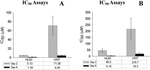
Figure 3. Effects of mevalonate (MLA) on proliferation in the presence or absence of lovastatin (lov). Cells were cultured for 6 days in the presence of lovastatin (50 µM) with or without mevalonate at the stated concentrations. Proliferation was assayed at day 6 by the MTS assay. (N = 3 for each cell line) (A) HL60 Cells (B) U937 cells.
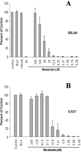
Figure 4. Number of days of statin exposure required for maximal statin-induced cytotoxicity. After various durations of lovastatin exposure, mevalonate was added to the cells to rescue them from the cytotoxic effects of statins in order to determine the minimum duration of statin exposure needed to induce a cytotoxic effect. (A) Duration of lovastatin exposure required at high concentrations (50 µM). HL60 and U937 cells were cultured with 50 M lovastatin for 0, 1, 2, 4, 7, 24 or 48 h prior to the addition of 200 µM Mevalonate. The culture was then incubated for 4 additional days and then assayed using the MTS-based proliferation assay (N ≥ 3 for each cell line). (B and C): IC50 determinations by day of rescue. Mevalonate was added on the indicated day of lovastatin treatment. IC50 values were determined on day 6 (N = 3 for each cell line). (B) HL60 Cells. (C) U937 Cells.
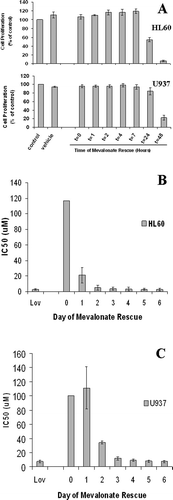
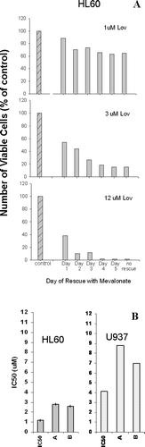
Figure 5. Effect of lovastatin pre-treatment on viability at clinically attainable concentrations. (A) Viability as assessed by Trypan blue staining: HL60 cells were treated with 1, 3 or 12 µM lovastatin on Day 0. Mevalonate (200 µM) was added to the cells on day 1, 2, 3, 4 or 5. The effects on viability determined on day 6 as measured by cell counts and Trypan blue uptake. N = 2; one example is shown. (B) Ability to recover from lovastatin after 72 h of exposure. HL60 or U937 cells were exposed to lovastatin for 72 h and then rescued with mevalonate (200 µM). They were allowed to proliferate for an additional 6 days. The IC50 was determined after a total of 3 days of lovastatin exposure and 6 days of mevalonate-rescued growth, i.e. on day 9. Two separate assays were performed and the results for each are as shown (N = 2 for each cell line, with the individual samples indicated by “A” or “B”). For comparison purposes, the IC50 values as determined previously are plotted (same data as , day 6).