
Figures & data
Table 1. Associations between the expression of PD-1, PD-L1, and PD-L2 in PTLD tissue, PTLD subtype, and clinical features.
Figure 1. Proportion of PTLDs positive for PD-1 in tumor-infiltrating cells (A) and for PD-L1 and PD-L2 in tumor cells (B).
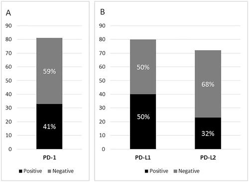
Figure 2. Expression of PD-1, -L1, and -L2 by subtype and EBV-status of PTLD.
PTLD: posttransplant lymphoproliferative disorder; EBER: Epstein–Barr virus-encoded RNA; LMP1: latent membrane protein 1; PD-1: programed death protein 1; PD-L1: programed death ligand 1; PD-L2: programed death ligand 2; H: heart; K: kidney; LU: lung; P: pancreas; LI: liver; DLBCL: diffuse large B-cell lymphoma; NGC: non-germinal center; GC: germinal center; Aggressive B: aggressive B-cell lymphoma, unclassifiable; Aggressive T: aggressive T-cell lymphoma, unclassifiable; ALCL: anaplastic large cell lymphoma; EATL: enteropathy-associated T-cell lymphoma; HSTL: hepatosplenic T-cell lymphoma; LBTL: lymphoblastic lymphoma; PTCL-NOS: peripheral T-cell lymphoma, unspecified; L-UNC: lymphoma, unclassifiable; +: positive; –: negative; MD: missing data.
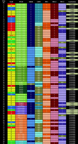
Figure 3. Immunohistochemical stainings of PD-1, PD-L1, and PD-L2 in PTLD. First row: Anaplastic large cell lymphoma (ALCL) positive in all three stainings, with a membranous staining pattern for PD-1(A) and PD-L1 (B), and a cytoplasmic and nuclear pattern for PD-L2 (C); Second row: Diffuse large B-cell lymphoma (DLBCL) of non-germinal center (non-GC) type with a membranous staining pattern for PD-1 (D) and PD-L1 (E), but negative for PD-L2 (F); Third row: DLBCL of GC type positive for PD-1 in a membranous pattern (G), with <5% of the tumor cells positive for PD-L1, therefore regarded as PD-L1 negative (H), but PD-L2 positive in a cytoplasmic and nuclear pattern (I); Fourth row: DLBCL of non-GC type negative for PD-1 (J), but positive for PD-L1 in a membranous (K) and PD-L2 in a cytoplasmic pattern (L).
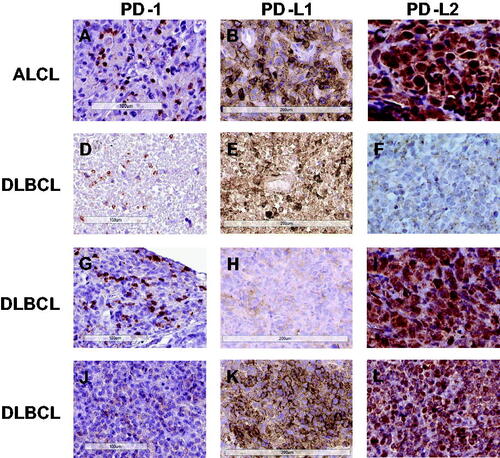
Table 2. Expression of PD-1, PD-L1, and PD-L2 in PTLD tissue.
Table 3. Combinations of expression of PD-1, PD-L1, and PD-L2 by subtype and EBV-status of PTLD.