Figures & data
Figure 1. Response to very low-dose radiotherapy. (A,B) Coronal (A) and axial (B) images demonstrating the radiation plan for a 67-year-old woman with stage IE BALT lymphoma of the left upper lobe treated with 4 Gy in 1 fraction. Color wash shows radiation dose. (C,D) PET/CT images in a 64-year-old man with stage IE BALT lymphoma of the right upper lobe (RUL) before (A, SUV 3.8) and 2 months after treatment with 4 Gy in 2 fractions (B, SUV 1.2). (E,F) PET/CT images in a 60-year-old woman with recurrent BALT lymphoma of the RUL before (C, SUV 10.1) and 6 months after treatment with 4 Gy in 2 fractions (D, SUV 1.9). (G,H). PET/CT images for the patient shown in (A, B) before (A, SUV 1.6) and 3 months after radiotherapy (F, SUV 0.6).
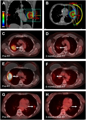
Table 1. Patient characteristics.
Table 2. Treatment outcomes.
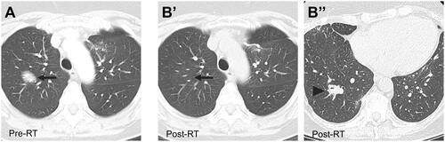
Figure 2. Regional Failure after radiotherapy for BALT lymphoma. (A) Axial CT image in a 48-year-old woman with recurrent BALT lymphoma of the RUL before treatment with 30 Gy in 15 fractions. (B) Axial CT images showing a CR in the treated lesion (arrow, B’) and regional recurrence in the right lower lobe (arrowhead, B’’) 17 months after radiotherapy.