Figures & data
Table 1. Description of digital pulse wave analysis (DPA) variables and effects of physiological conditions.
Table 2. Demographic data of the study population.
Table 3. Associations at baseline measurements between DPA variables and maternal age, heart rate, logarithmic value of ovarian sensitivity index (logOSI) and grade of ovarian response at controlled ovarian hyperstimulation.
Table 4. Hemodynamic effects of oocyte stimulation and pregnancy, compared with basal measurements before conception. For details, see text. Digital pulse wave analysis values are indices without quantitative measures.
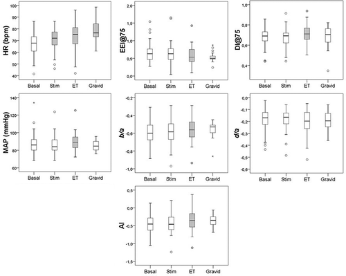
Figure 1. Boxplots showing cardiovascular changes after administration of follicle-stimulation hormone (Stim), at embryo transfer (ET) and in early pregnancy (Gravid) compared to baseline values (Basal). Gray boxes indicate significant changes (p < 0.05) compared with basal measurements, and star symbols denote values outside the panel boxes. HR, heart rate; MAP, mean arterial pressure; AI, aging index; EEI@75, cardiac ejection elasticity index adjusted to heart rate 75 bpm; DI@75, dicrotic index adjusted to heart rate 75 bpm.