Figures & data
Figure 1. The surface morphology and fiber diameter distribution of poly(ε-caprolactone) (a,b), PCL/Calophyllum inophyllum oil (CIO)−2.5 (c,d), PCL/CIO−5 (e,f) and PCL/CIO−7.5 (g,h) fiber mats, respectively.
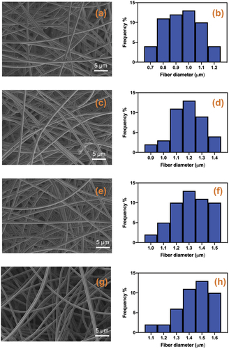
Table 1. Sample name, average fibre diameter (μm) and contact angle values of electrospun fibre mats.
Figure 2. FT-IR spectrums of pure poly(ε-caprolactone) (PCL), Calophyllum inophyllum essential oil loaded PCL fiber mat (PCL/CIO) and Calophyllum inophyllum (CIO) essential oil.
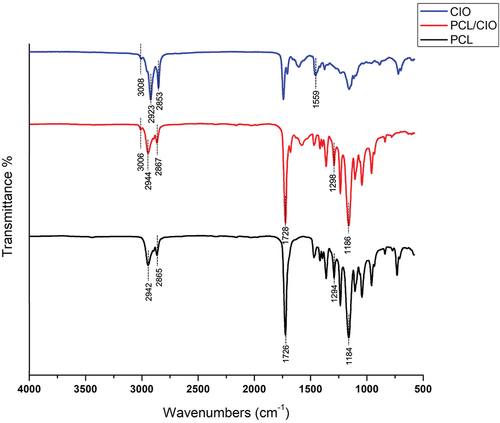
Figure 3. Swelling degree (%) of pure poly(ε-caprolactone) (PCL) fiber mat and Calophyllum inophyllum essential oil loaded PCL fiber mats (PCL/CIO), PCL/CIO−2.5, PCL/CIO−5 and PCL/CIO−7.5.
Figure 4. Cumulative release of Calophyllum inophyllum oil (CIO) from poly(ε-caprolactone) (PCL)/CIO fiber mats.
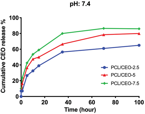
Figure 5. Anti-bacterial activity of poly(ε-caprolactone) (PCL)/CIO fiber mats against Staphylococcus aureus (a) and Escherichia coli (b). *p<0.05, **p<0.01 compared to the control.
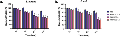
Figure 6. In vitro cytotoxicity of poly(ε-caprolactone) (PCL) electrospun fiber mats loaded with different concentrations of Calophyllum inophyllum oil (CIO) (PCL/CIO−2.5, PCL/CIO−5, and PCL/CIO−7.5) on NHDF cells.
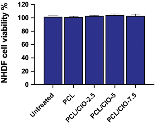
Figure 7. In vitro wound healing activity of poly(ε-caprolactone) (PCL)/CIO fiber mats on normal human dermal fibroblast cells.
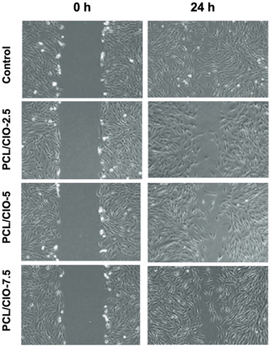
Figure 8. Total antioxidant (a) and oxidant capacities of poly(ε-caprolactone) (PCL)/CIO fiber mats. *p<0.05 compared to the control.
Figure 9. Cytotoxic activity of poly(ε-caprolactone) (PCL)/CIO fiber mats on macrophages (a). Interleukin−1β levels of poly(ε-caprolactone) (PCL)/CIO fiber mat treated cells (b).