
Figures & data
Figure 1. Visible observation of silver nanoparticles.
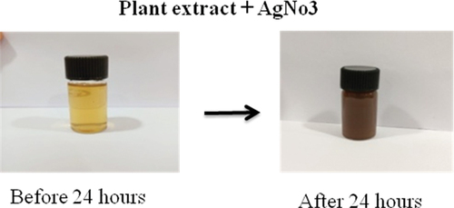
Figure 2. UV- Visible spectrum of silver nanoparticles with SPR band at 450 nm.
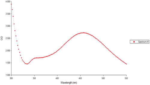
Figure 3. FTIR spectroscopic analysis of silver nanoparticles.
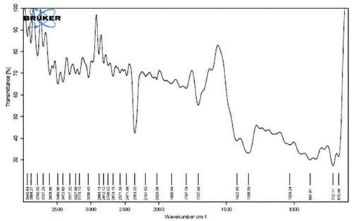
Figure 4. XRD of silver nanoparticles.
Figure 5. a) SEM and b) EDAX of silver nanoparticles.
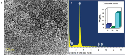
Figure 6. TEM of synthesized silver nanoparticles.
Figure 7. Antibacterial activity of plant extract on different microorganisms.
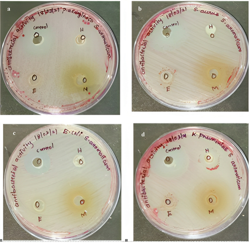
Figure 8. Antibacterial activity of silver nanoparticles.
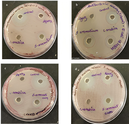
Table 1. Antibacterial activity of synthesized silver nanoparticles.
Table 2. Antibacterial activity of plant extracts.
Figure 9. Antimycobacterial activity of silver nanoparticles and plant extract.
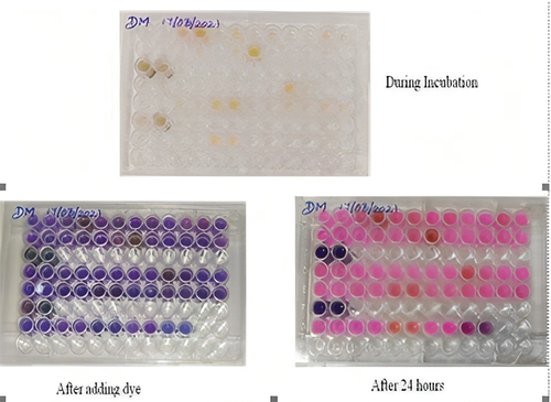
Table 3. Quantitative analysis on antimycobacterial activity and plant extract.
Figure 10. Zebra fish embryo – morphological change was noticed with high concentration of nanodrug.
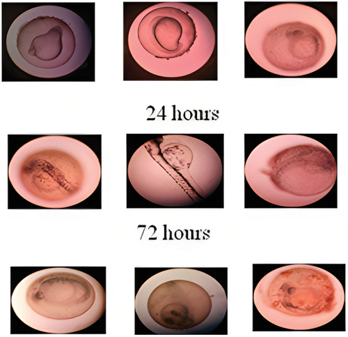
Table 4. The cytotoxicity assay of plant extract Syzygium aromaticum was assessed by zebra fish embryo.
Figure 11. Survival rate and LC50 concentration of Artemia salina for silver nanoparticle and plant extract.
Data availability statement
The authors confirm that the data supporting the findings of this study are available within the article.