

Abstract
As T cells transit between blood, lymphoid organs, and peripheral tissues, they experience varied levels of oxygen/hypoxia in inflamed tissues, skin, intestinal lining, and secondary lymphoid organs. Critical illness among COVID-19 patients is also associated with transient hypoxia and attenuation of T cell responses. Hypoxia is the fulcrum of altered metabolism, impaired functions, and cessation of growth of a subset of T cells. However, the restoration of normal T cell functions following transient hypoxia and kinetics of their phenotype-redistribution is not completely understood. Here, we sought to understand kinetics and reversibility of dichotomous T cell responses under sustained and transient hypoxia. We found that a subset of activated T cells accumulated as lymphoblasts under hypoxia. Further, T cells showed the normal expression of activation markers CD25 and CD69 and inflammatory cytokine secretion but a subset exhibited delayed cell proliferation under hypoxia. Increased levels of reactive oxygen species (ROS) in cytosol and mitochondria were seen during dichotomous and reversible attenuation of T cell response under hypoxia. Cell cycle analysis revealed maximum levels of cytosolic and mitochondrial ROS in dividing T cells (in S, G2, or M phase). Hypoxic T cells also showed specific attenuation of activation induced memory phenotype conversion without affecting naïve and activated T cells. Hypoxia-related attenuation of T cell proliferation was also found to be reversible in an allogeneic leukocyte specific mixed lymphocyte reaction assay. In summary, our results show that hypoxia induces a reversible delay in proliferation of a subset of T cells which is associated with obliteration of memory phenotype and specific increase in cytosolic/mitochondrial ROS levels in actively dividing subpopulation. Thus, the transient reoxygenation of hypoxic patients may restore normal T cell responses.
Graphical Abstract
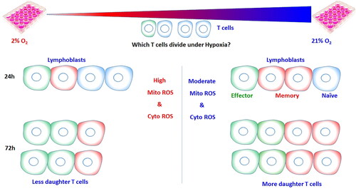
Activated T cells cultured under hypoxic conditions show increased cellular ROS and mitochondrial ROS levels and there is limited expansion of memory phenotype T cells under as compared to activated T cells cultured under normoxia.
Acknowledgments
The authors acknowledge the technical assistance provided by Ms. Binita K. Kumar, Mr. D. V. Kathole, and Mr. B. A. Naidu.
Author contributions
D.K.M., D.S., and S.S.K. contributed to the conception and design of the experiments. D.K.M. was responsible for conducting experiments. D.K.M. and D.S. were responsible for data analysis and interpretation. D.K.M. drafted the manuscript, D.S. corrected and edited the manuscript whereas S.S.K. finalized the manuscript.
Disclosure statement
No potential conflict of interest was reported by the author(s).