Figures & data
FIG. 1 Photomicrographs of freshly prepared APAmicrocapsule thalidomide formulation (size 300 μm ± 50 μm, ×250).
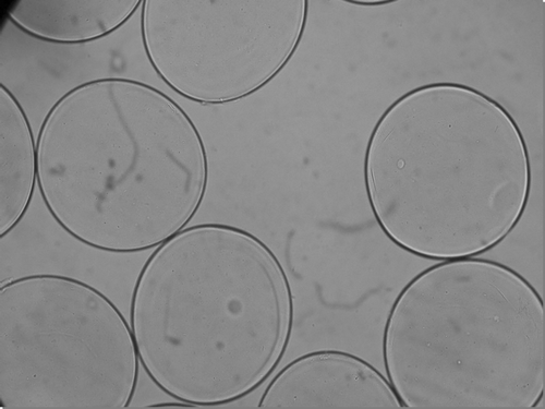
FIG. 2 Standard curve for TNF-α secretion from RAW 264.7 macrophage cells.
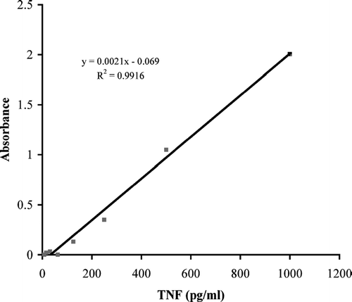
FIG. 3 The concentration of TNF-α secretion from RAW 264.7 macrophage cells stimulated with 10 μ g/ml of LPS. These values are compared with TNF-α secretion from stimulated RAW 264.7 macrophage cells in the presence of APA encapsulated thalidomide. Comparisons were made after incubation times of 0.5, 1, 3, 12, 24, and 48 hr.
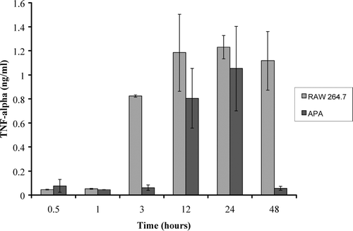
FIG. 4 The concentration of TNF-α secretion from RAW 264.7 macrophage cells stimulated with 100 μ g/ml of LPS. These values are compared with TNF-α secretion from stimulated 264.7 macrophage cells in the presence of APA encapsulated thalidomide. Comparisons were made after incubation times of 0.5, 1, 3, 12, 24, and 48 hr.
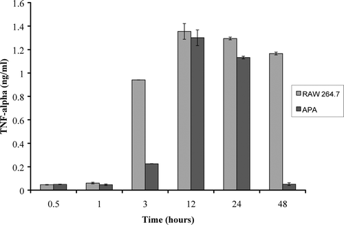
FIG. 5 TNF-α secretion from RAW 264.7 macrophage cells in comparison to RAW 264.7 macrophage cells incubated with APA encapsulated thalidomide at time intervals from 0.5, 1, 3, 12, 24, and 48 hr. Turquoise and blue lines in the graph represent cells stimulated with 100 μ g/ml of LPS versus the green and yellow lines that represent cells stimulated with 10 μ g/ml of LPS.
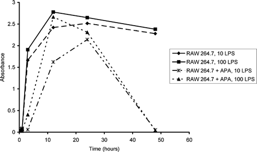
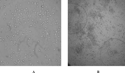
FIG. 6 Photomicrographs of RAW 264.7 control macrophage cells and experimental RAW 264.7 macrophage cells after exposure to APA encapsulated thalidomide formulations. (A) Photomicrograph of control macrophages (×200) (B) Experimental: photomicrograph of macrophages after 48 hr exposure to APA microcapsules thalidomide exposers (×200). APA microcapsule thalidomide formulations also are seen in the photomicrograph.