Figures & data
FIG. 1 FTIR spectra of (a) chitosan phthalate microspheres and (b) chitosan microspheres.
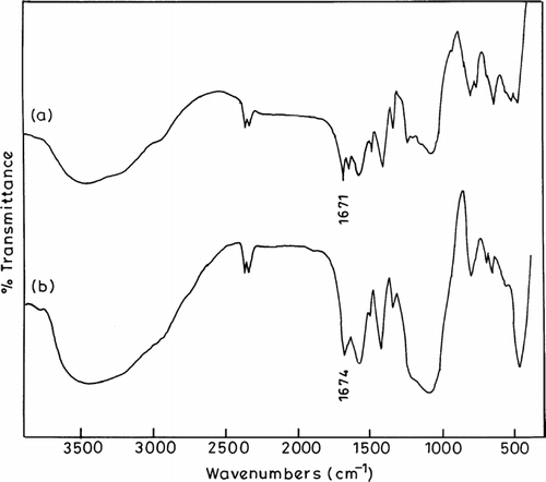
FIG. 2 Photomicroscopy of chitosan phthalate microspheres.
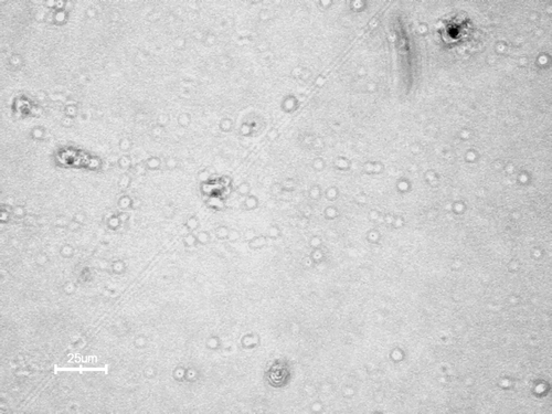
FIG. 3 SDS-polyacrylamide gel electrophoresis (SDS-PAGE) of insulin. Lane 1: supernatant plain microspheres; Lane 2: supernatant after insulin-loading process; Lane 3 and 4: released samples from the insulin-loaded microspheres in PBS (pH 7.4); Lane 5: insulin standard.
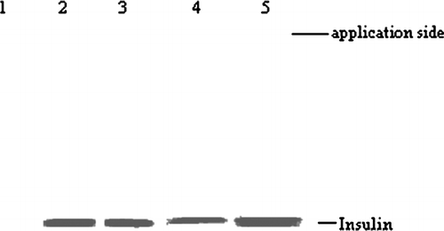
TABLE 1 Degradation of insulin and insulin-loaded chitosan phthalate microspheres in pepsin and trypsin solution
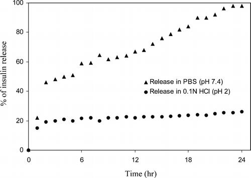
FIG. 4 In vitro release of insulin from microspheres in the presence of PBS (pH 7.4) and 0.1N HCl (pH 2.0).