Figures & data
TABLE 1 Mean diameter zeta potential and encapsulation efficiency of microparticles prepared by the solvent evaporation method
DIAG. 1. Representation of the typical psoralen (A) and psoralen A (3-ethoxy carbonyl-2H-benzofuro[3,2-f]-1-benzoyiran-2-one) (B).
![DIAG. 1. Representation of the typical psoralen (A) and psoralen A (3-ethoxy carbonyl-2H-benzofuro[3,2-f]-1-benzoyiran-2-one) (B).](/cms/asset/a33b6956-e0a4-4fdc-a997-e0b647d31f92/idrd_a_164015_uf0001_b.gif)
FIG. 1 Cumulative release profile of psoralen A from PLGA microparticle into buffer medium after 15 days.
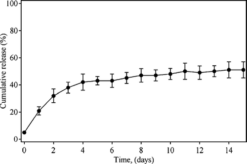
FIG. 2 Absorption spectrum of psoralen-loaded microparticle. The spectrum is deconvoluted into three components: (–)-deconvoluted absorption of psoralen A. Inset: (- - -)-glycerol and (—)-PLGA microparticle-psoralen in glycerol.
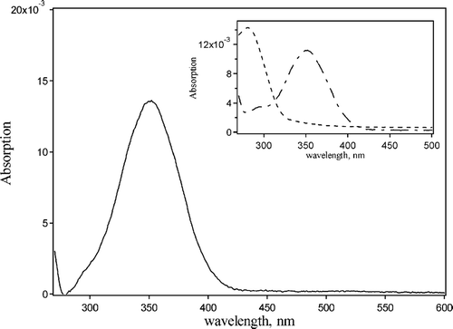
FIG. 3 Ultrastructural characterization of PLGA-microparticle loaded with psoralen A. (A) Representative SEM shows the surface of PLGA microparticle loaded with psoralen A (30000×). (B) PLGA-microparticle (SEM) (→)-phagocited by macrophage cell after incubation for 120 min (5000×). (C) Representative TEM of microparticle in incubation for 120 min peritoneal exudate cell population: neutrophil-Nϕ, eosinophil-Eo, hemacea-He, and selective phagocytose by macrophage-MC (3200×).
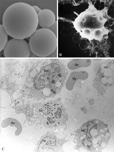
FIG. 4 Phagocytose of PLGA-microparticle loaded with psoralen A by macrophage peritoneal exudate. (A) PLGA microparticle (**) interacting with cellular membrane in the process of phagocytosis (15 min incubation). (B) Phagosome with PLGA microparticle (30 min incubation). (C) Psoralen A after microparticle digestion by macrophage (120 min incubation). (D) PLGA microparticle inside the macrophage cell, without irradiation (incubated for 120 min), mitochondria-Mi, lysosomes-Ly, nucleus (N), and granular endoplasmatic reticulum-Reg. (E) Photo-damage (PUVA) in macrophage cell with microparticle after 120 min incubation: cytoplasmic vesiculation, mitochondria condensation (Mi), and a swelling of granular endoplasmatic reticulum (Reg) and nuclear membrane (arrow) (12000×).
