Figures & data
FIG. 1 Photomicrograph shows TEM of elastic liposomes (× 30000).
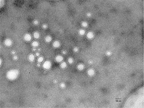
TABLE 1 Composition and characterization of elastic liposomal formulations
FIG. 2 (A) Fluorescent intensity (AU) versus skin depth (μm) studies reveal skin penetration profile of elastic liposomes. AU = arbitrary unit and (B) virtual channel-like structures can be visualized (yellow arrows represent the stained channels) at the skin depth of 10 μm.
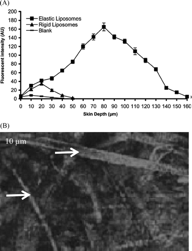
FIG. 3 Percentage cumulative drug release from elastic liposomal formulation, conventional liposomal formulation, and plain drug solution through albino rat skin (mean ± S.E., n = 3).
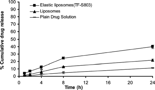
TABLE 2 Permeation parameters of acyclovir-loaded formulations across rat skin after 24 hours
FIG. 4 Drug plasma concentration profile after transdermal application of different formulations (mean ± S.E. n = 6).
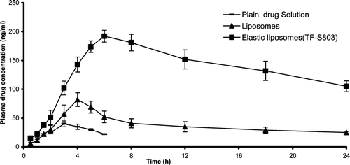
TABLE 3 Pharmacokinetic parameters of acyclovir