Figures & data
Table 1. Composition of gel pluronic® F127.
Table 2. Composition of Pluronic Lecitin Organogel (PLO).
Figure 1. Changes in viscosity (cP) of Pluronic® F-127 gel at 25°C with time.
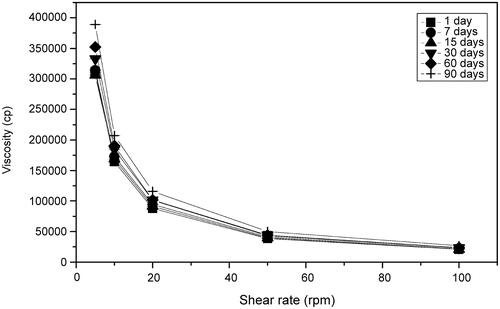
Figure 2. Changes in viscosity (cP) of Pluronic® F-127 gel at 4°C with time.
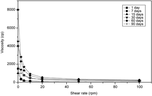
Figure 3. Changes in viscosity (cP) of Pluronic lecitin organogel at 4°C with time.
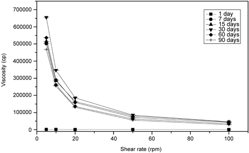
Figure 4. Changes in viscosity (cP) of Pluronic lecitin organogel at 25°C with time.
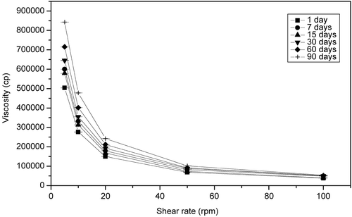
Figure 5. Photomicrograph of Pluronic gel 7 days after preparation.
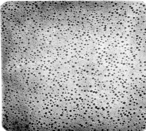
Figure 6. Photomicrograph of pluronic lecithin organogel 7 (a), 30 (b), and 90 (c) days after preparation.
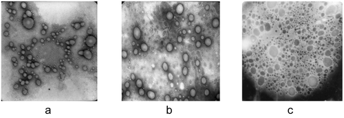
Table 3. Changes in micelle size in Pluronic F127® gel and in vesicle size in pluronic organogel formulations.
Figure 7. Photomicrograph of pluronic gel 1 year after preparation.
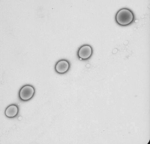
Figure 8. Photomicrograph of pluronic lecithin organogel 1 year after preparation.
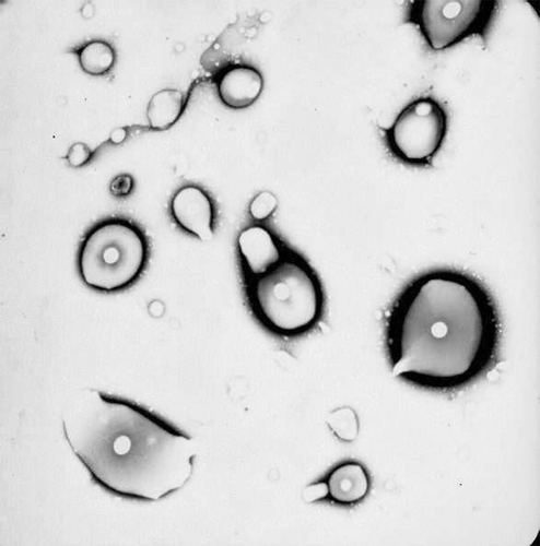
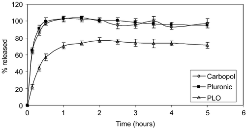
Figure 9. Percentage of drug released from different formulations in release device without membrane.
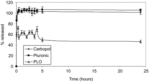
Figure 10. Percentage of drug released from different formulations in Franz-type cells.