Figures & data
Figure 1. Polyvinylalcohol partial nicotinic ester.
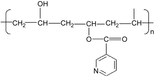
Figure 2. Mean diameter (nm) of the micelles formed at 37°C in water by dissolution, at different polymer concentrations, of PVA-NIC 5% (▵), PVA-NIC 8% (◊), PVA-NIC 15% (□), ATRA:PVA-NIC 5% (▴), ATRA:PVA-NIC 8% (♦), and ATRA:PVA-NIC 15% (▪). Each value represents the mean ± SD of four independent experiments.
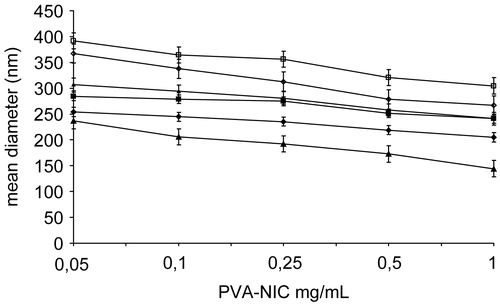
Figure 3. Fractional release (%) of ATRA from micellar complexes in PBS at 37°C: ATRA:PVA-NIC 5% (▴), ATRA:PVA-NIC 8% (♦), and ATRA:PVA-NIC 15% (▪). Results are the mean ± standard error from four independent experiments.
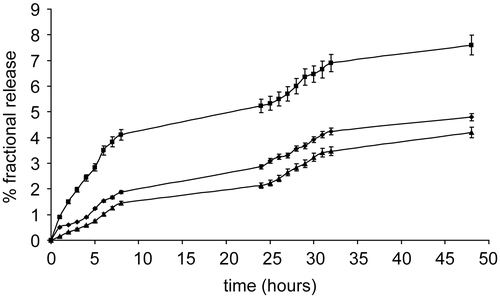
Figure 4. Time course of the growth inhibiting and cytotoxic properties of pure ATRA (5 μM), complexed ATRA (5 μM), and pure polymer (PVA-NIC 15%, 25 μM) on human neuroblastoma LAN-5 cells. * p < 0.02; ** p < 0.001; *** p < 0.0001. White columns: control; black columns: pure polymer; gray columns: pure ATRA; dark gray columns: complexed ATRA.
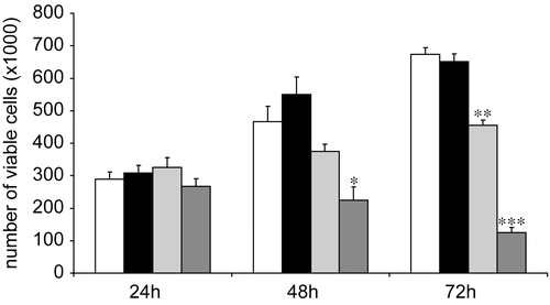
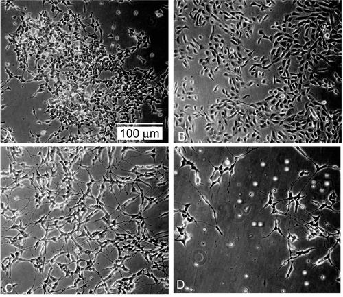
Figure 5. Morphologic appearance of LAN-5 cell cultures after 72 h of treatment. (A) control; (B) pure polymer (36 μM); (C) pure ATRA (5 μM); (D) complexed ATRA (5 μM). Bar = 100 μm. One representative experiment out of three performed is shown.