Figures & data
Figure 1. Schematic representation of the preparation process of the magnetic chitosan–MTX microspheres.
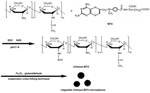
Figure 2. A typical SEM image of magnetite chitosan–MTX microspheres.
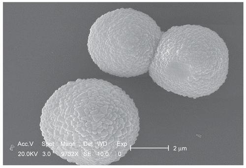
Figure 3. FTIR spectra of chitosan (a), MTX (b), Chitosan–MTX (c), and magnetite chitosan–MTX microspheres (d).
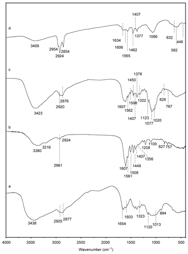
Table 1. Assignments of the characteristic FTIR absorption bands presented in .
Figure 4. XRD patterns of the prepared magnetite nanoparticles (a) and magnetite chitosan–MTX microspheres (b).
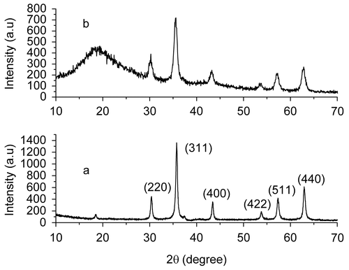
Figure 5. TGA curves of the prepared magnetite nanoparticles (a) and magnetite chitosan–MTX microspheres (b).
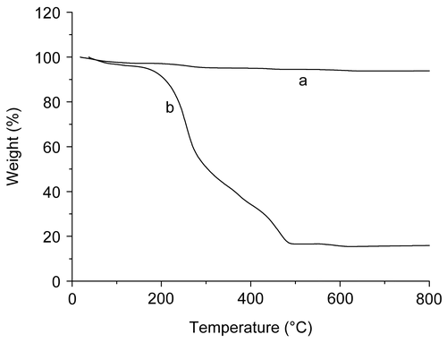
Figure 6. Release profiles of methotrexate from magnetic chitosan–MTX microspheres in the presence of 0.1 mg/ml crude protease from bovine pancreas with various pH values at 37°C.
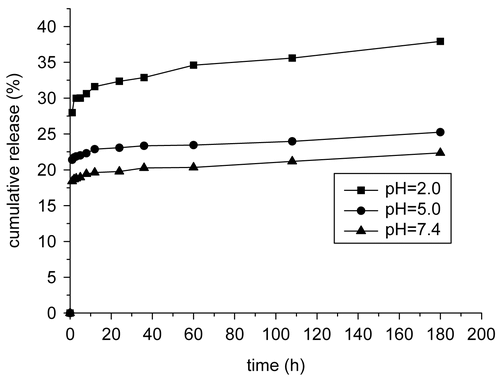
Figure 7. Release profiles of methotrexate from magnetic chitosan–MTX microspheres (a) and chitosan-MTX conjugations (b) at pH = 7.4 in the presence of 0.1 mg/ml crude protease from bovine pancreas at 37°C.
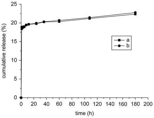
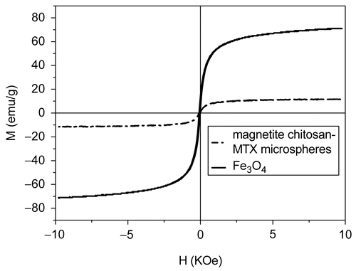
Figure 8. Magnetization curves for the prepared magnetite nanoparticles (solid) and magnetite chitosan-MTX microspheres (dash dot) at room temperature.