Figures & data
Figure 1. The principles of RED and iontophoretic drug delivery (A) the transport of cations and anions during the controlled mixing of saltwater and freshwater through selective ion exchange membranes (B) the net flow of drug solution from the anode to the cathode (or from the cathode to the anode according to charge of drug solution) under the influence of an electric current. AEM: anion exchange membrane; CEM: cation exchange membrane.
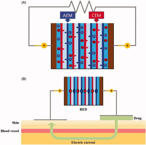
Figure 2. (A) Schematic of the RED system and experimental set up and (B) appearance of the disposable RED system.
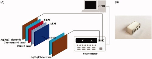
Figure 3. Polarization and power curves of the RED. Ncell = 8 and C = 0.01 M.
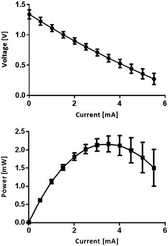
Figure 4. Time–current curves of the RED. Ncell = 8 and C = 0.01 M.
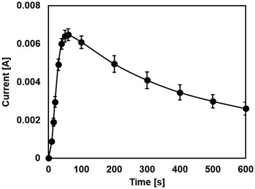
Figure 5. Distribution of FITC-PLL solution topically applied with or without RED on porcine skin. All images in this figure are magnified at 200×. Control, no treatment; passive diffusion, only topical application for 30 min and 1 h, respectively; RED, iontophoretic drug delivery for 30 min and 1 h, respectively (Left column, differential interference contrast (DIC) images; middle column, fluorescence images; right column, merged images).
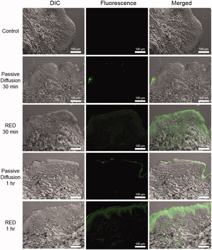
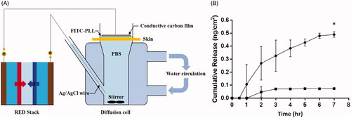
Figure 6. (A) Schematic of Franz diffusion cell apparatus (B) In vitro penetration of FITC-conjugated-PLL using a Franz diffusion cell equipped with micro pig skin with or without RED iontophoresis for 1 h. (▪; Passive diffusion, n = 3, •; RED iontophoresis, n = 2).