Figures & data
Figure 1. BPD preparation, intra-testicular injection, and foreign gene expression after TMGT in the testes. (A) The structure of pAcGFP-N1 plasmid as well as the loci of primers (red arrows) designed for PCR procedure. (B, C) Surgical procedures for intra-testicular injection, in which a 27-gauge needle is used to pierce the tunica albuginea of the testis at one of the ends of the long axis to assist penetration of mouth pipette into the tissue (B). Mouth pipette was withdrawn from the testis bit by bit while solution was injected into the testes (C). (D) The clusters of BMPs (red arrow) that have conjugated with PEI-DNAs (red arrow head) observed under TEM. The size distribution of BMPs ranged from 45 to 55 nm. Amplification: × 60 000. (E, F) BPDs were presented within the cells of seminiferous tubule (red arrows). BPDs were observed both within the cytoplasm as well as nuclear of spermatogenic cells (E, bar = 200 nm) and within the tails of sperms (F, bar = 500 nm). (G, H, I) In vivo and ex vivo GFP fluorescence detection. (G) The fluorescence of GFP protein was presented in the testes of BMP-PEI group mouse under KODAK Image Station In-Vivo FX system. (H) No fluorescence was observed in the testes of mice coming from both groups, as compared with the GFP transgene positive mice obtained through conventional pronuclear injection method (black mouse on the right), but can be detected under IVIS QUANTUM FX system when mouse testes were recovered. (J) The expression of GFP protein was shown in testes injected with BPDs and Lipo-DNAs (red arrow) under fluorescence microscope.
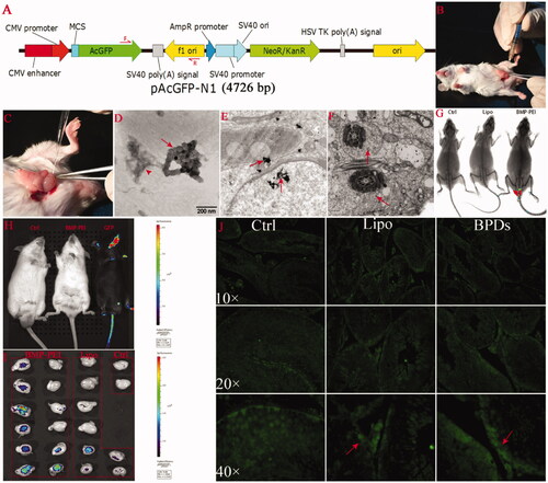
Figure 2. The influence of different DNA carriers on sperm motility after TMGT. From chart A through D, different letters in each chart indicate significant differences among groups (ANOVA, p < 0.05). As can be seen, almost all sperm motility parameters were significantly different among BPDs, Lipo-DNAs, and the controls except ALH (B). In addition, the increase of VAP, VSL, VCL, LIN, and STR in BPD groups, which represents the improved sperm motility, was significantly higher than the Lipo-DNA groups.
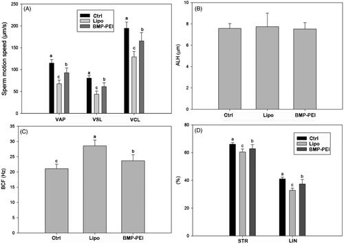
Figure 3. Genotype analysis of the first- and the second generations of founder mice derived from TMGT. (A, B) Demonstration of PCR analysis of plasmid encoding GFP integration in the chromosomes of offspring derived from some of the founder mice tail tissue. A: BPDs; B: liposome. (C) The ratios of heterozygous in the first filial between BPD and Lipo-DNA treatments, of which the ratio in BPDs was 6 times higher than the Lipo-DNA group (73.8% versus 11.6%, p < 0.05). (D) Demonstration of the genotypes of the second generation of founder mice after BPD injection. It showed that transgene can be successfully inherited by the second filial after inbreeding of the first filial. (E) Demonstration of the transgene that was expressed in some of the tissues of the second filial bodies after inbreeding of the first filials, as was approved by Kodak Image Station In-Vivo FX system. These transgene mice came from the founder mice No. 1.
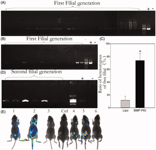
Figure 4. The efficiency of TMGT between BPD and Lipo-DNA groups. (A) The ratios of founder mice obtained from different treatments were significantly different from each other, in which founder mice in BPDs were 3 times more than that in Lipo-DNA group. (B) Upon litter size of founders, there was no significant difference between the two groups. * Indicated significant different (p < 0.05).
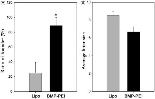
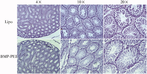
Figure 5. Histochemistry evaluation of the structures of testes that performed TMGT. The results showed that no obvious histological destructions based on seminiferous tubules was observed in the testes of both BPD and Lipo-DNA groups.