Figures & data
Figure 1. Overview of the pNIPAM hydrogel IO. Schematic of the self-reporting feature of the pNIPAM hydrogel IO particles during drug release (green circles). The accompanying blue shift in the reflection peak of a particle at triggered release is indicated by the two wavelengths λ1 and λ2 (Zhang et al., Citation2015a).
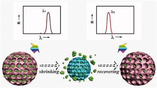
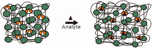
Figure 2. PC sensing materials that consist of CCA surrounded by a polymer hydrogel. In this example, the hydrogel volume swells in response to the interaction between glucose and the molecular recognition element (Alexeev et al., Citation2005).