Figures & data
Figure 1. Effect of 2OHOA content on DSC thermograms of DMPC liposomes. Liposomes were prepared with the indicated molar ratio of DMPC and 2OHOA.
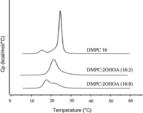
Figure 2. Effects of 2OHOA insertion on the physicochemical characteristics of liposomes. (A) Effects of 2OHOA content on the mean particle size and zeta potential of liposomes. Liposomes were prepared with the indicated molar ratio of DMPC and 2OHOA. For comparison, oleic acid-inserted liposomes were prepared with a 16:8 molar ratio of DMPC and oleic acid. (B) Effect of 2OHOA insertion on the mean size of liposomes prepared with DOPC or DPPC. Liposomes were prepared with a 16:8 molar ratio of PC and 2OHOA. (C) Representative TEM image of 2OHOA-inserted liposomes. Liposomes were prepared with a 16:8 molar ratio of DMPC:2OHOA. (D) Time-dependent changes in the mean particle size of liposomes with varying 2OHOA content. Liposomes prepared with the indicated ratio of DMPC and 2OHOA were stored at room temperature for up to 28 days. Data are expressed as means ± SD (n = 3; *p < .05, **p < .005 compared with the initial condition).
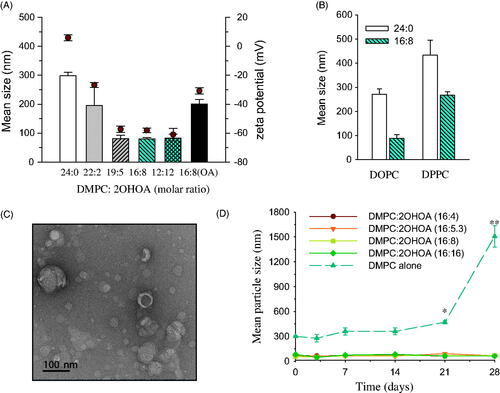
Figure 3. Effects of liposomal insertion on the anticancer activity of 2OHOA. (A) Comparison of the anticancer efficacy of free and liposome-inserted 2OHOA. Cell growth and viability were determined by MTT assays after incubating UM-SCC-1 tumor cells with different formulations for 72 h. The free 2OHOA solution was prepared by dissolving 2OHOA in DMSO, and liposomes were prepared with DMPC alone or a 16:8 molar ratio of DMPC:2OHOA. Liposomes composed of DMPC alone were added to cells after preparing dilutions corresponding to the dilution of DMPC:2OHOA liposomes. (B) Effects of liposome-inserted 2OHOA on the growth and viability of cancer cells and normal cells. Cancer cells (UM-SCC-1) and normal cells (MRC5) were incubated with different dilutions of DMPC:2OHOA (molar ratio, 16:8) liposomes for 72 h, after which MTT assays were performed. (C) Effect of DMPC supplementation on the anticancer activity of 2OHOA. UM-SCC1 cells were incubated with 120 μM 2OHOA (DMSO solution) in the presence of the indicated concentration of DMPC (DMSO solution) for 48 h, after which MTT assays were performed. Results are expressed as percentage growth (means ± S.D. of triplicate wells) relative to control cells (*p < .05, **p < .005 compared with 2OHOA treatment alone).
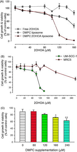
Figure 4. Effects of 2OHOA insertion on the drug-incorporation capacity of liposomes. Effects of 2OHOA insertion on the liposome-incorporated concentration of (A) mitoxantrone, (B) paclitaxel, and (C) ATRA. (A and B) Liposomes were prepared with DMPC alone (24 μmole/ml) or DMPC:2OHOA (molar ratio, 16:8; total, 24 μmole/ml) together with 0.25 mg of mitoxantrone or 1.5 mg of paclitaxel. (C) Liposomes were prepared with DMPC:CHOL (molar ratio, 24:2; total, 26 μmole/ml) or DMPC:2OHOA:CHOL (molar ratio, 12:12:2; total, 26 μmole/ml) containing 2 mg of ATRA. Data are expressed as means ± SD (n = 3; **p < .005, ***p < .0001 compared with 2OHOA-free liposomes). (D) Schematic depiction of the structure of 2OHOA-inserted liposomes. (E) Effect of 2OHOA insertion on the time-dependent leakage of ATRA incorporated in liposomes. Liposomes were prepared as described in (C) and stored at 27 °C up to 48 h.
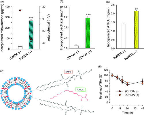
Figure 5. Effect of 2OHOA on the in vitro anticancer activity of ATRA. The growth-inhibitory effect of ATRA in mouse B16–F10 melanoma cells was evaluated by MTT assays after a 72-h incubation. (A) Growth-inhibitory effects of free ATRA in the presence or absence of free 2OHOA (150 μM). (B) Comparison of the growth-inhibitory effects of ATRA incorporated in conventional liposomes or 2OHOA-inserted liposomes. Liposomes used in the study were composed of DMPC:CHOL:ATRA (molar ratio, 24:2:0.53) and DMPC:2OHOA:CHOL:ATRA (molar ratio, 12:12:2:0.53). Data are expressed as percentage growth (means ± S.D. of triplicate wells) relative to control cells.
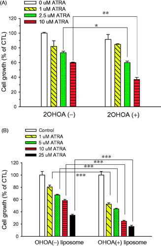
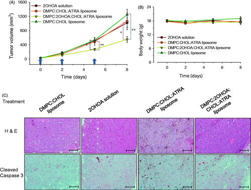
Figure 6. In vivo antitumor study. (A) Tumor growth and (B) body weight changes were monitored in a B16–F10 syngeneic mouse tumor model after treatment with free 2OHOA, ATRA-incorporated conventional liposomes, ATRA-incorporated/2OHOA-inserted liposomes, or empty conventional liposomes. Upward arrows indicate treatment time points. Data are presented as means ± SEM (n = 6–7; *p < .05, **p < .005). (C) Representative immunohistochemical images of syngeneic B16–F10 tumors obtained from each group of mice after treatment. Scale bars: 100 μm. The 2OHOA solution for treatment was prepared by dissolving 2OHOA at 6.4 mg/ml in distilled water containing 0.25% hydroxypropylmethylcellulose (Shin-Etsu Co., Tokyo, Japan) and 0.2% Tween-80 (Sigma-Aldrich Inc., St. Louis, MO). Empty conventional liposomes were composed of a 40:3 molar ratio of DMPC:CHOL. The composition of ATRA-incorporated conventional liposomes and ATRA-incorporated/2OHOA-inserted liposomes used were DMPC:CHOL:ATRA (molar ratio, 24:1.9:0.85) and DMPC:2OHOA:CHOL:ATRA (molar ratio, 12:12:1.9:0.78), respectively. The arrow indicates the active caspase-3 fragment; the brown-black pigments are melanin.