Figures & data
Figure 1. Scheme of potential bio-function of TPGS-based DDS for the treatment of cancer.
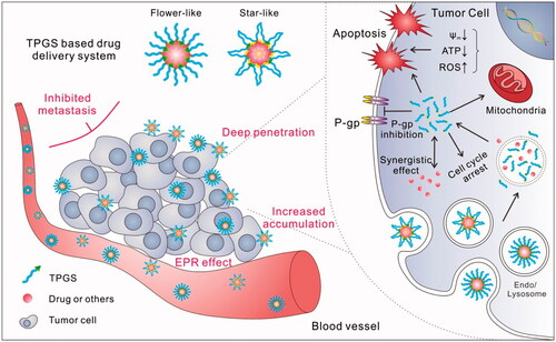
Figure 2. TPGS improves drug accumulation and penetration in tumors. (A) In vivo imaging, (B) Ex vivo imaging of excised tumors and (C) Intratumoral distributions of Did-loaded micelles in B16F10 tumor bearing mice. 1, Saline; 2, Did; 3, P84-TPGS mixed micelles; 4, F127 -TPGS mixed micelles; 5, F127 micelles (Cao et al., Citation2016).
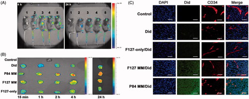
Figure 3. Mixed micelle-based complex NPs for the co-delivery of chemo-drug and shRNA. (A) Schematic illustration of NP preparation. (B) Anticancer effects and body weight changes of different treatments in A549/T bearing nude mice. (C) In vivo biodistribution of different formulations of PTX or shSur in different formulation at 0.5, 4, 12, and 24 h in A549/T bearing nude mice (Shen et al., Citation2014a).
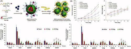
Figure 4. TNO3 and TPGS-S-S-PTX hybrid micelles. (A) In vivo tumor vascular permeability and blood perfusion presented by Representative MRI images in an S180 tumor model. (B) CLSM images for of micelle uptake and NO release in MCF-7/ADR cells. NO was detected by DAF-FM DA (green). Micelles were labeled by with RhB (red), and nuclei were stained by with DAPI (blue). Scale bar, 50 μm. (C) Representative immunofluorescentce images of blood vessels and tumor apoptosis of in MCF-7/ADR tumors. Blood vessels, nuclei and apoptotic cells were stained by α-CD31 antibody (red), DAPI (blue) and TUNEL (green), respectively (Yin et al., Citation2017b) (colour figure online).
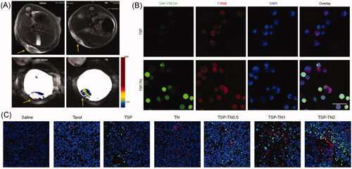
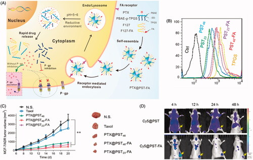
Figure 5. Redox/pH dual-sensitive PBAE-g-TPGS hybrid micelles. (A) Scheme of the targeting delivery and overcoming MDR. (B) Rh123 retention in MCF-7/ADR cells. (C) Live images of MCF-7/ADR tumor-bearing mice that were i.v. administered Cy5 loaded micelles. (D) In vivo anticancer activities (Yin et al., Citation2017a).