Figures & data
Table 1. Niosomes: (A) composition of the niosomes; (B) encapsulation efficiency of niosomes (F1–F6). (A)
Figure 1. Skin wounds (2 cm in diameter) on one side of the thorax of the dog.
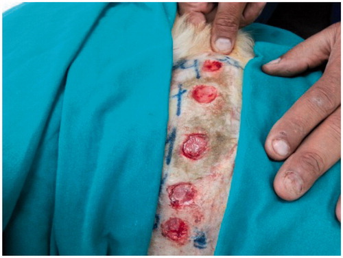
Figure 2. Phytochemical analysis. (A) Analysis of the extracts (ASE®100) using a Shimadzu spectrophotometer for total hypericins and HPLC-DAD for all other compounds. Hyperoside and isoquercitrin, being similar, were not separated under the present chromatographic conditions. MeOH: methanol; rt: room temperature; EtOH: ethanol. (B) HPLC-DAD chromatographic profile of the 80% ethanol extract at 70 °C (HE) using DIG-MAZ. 1, chlorogenic acid isomer at Rt 6.02; 2, chlorogenic acid at Rt 7.91; 3, rutin at Rt 16.48; 4, hyperoside at Rt 17.04; 5, isoquercitrin at Rt 17.25; 8, quercitrin at Rt 18.46; 9, quercetin at Rt 21.16; 10, I3’, II8 biapigenin at Rt 22.79; 11, pseudohypericin at Rt 26.02; 12, hyperforin analog at Rt 35.64; 13, hyperforin at Rt 36.54; adhyperforin at Rt 37.18.
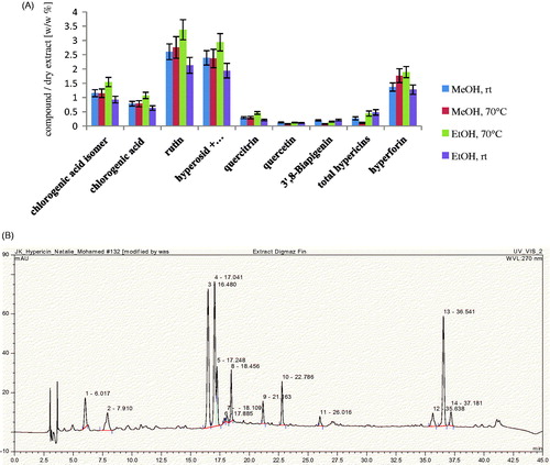
Figure 3. TEM micrographs of optimum niosomal formula (F1).
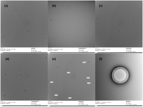
Figure 4. Release of total hypericins from niosomal gels containing 1.5% NaCMC and 3% HEC as polymers.
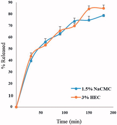
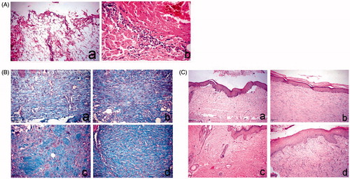
Figure 5. Histopathology of skin wound of dog. (A) At day 0 stained with H&E. a, large wound gap with complete loss of epidermal and dermal cell layer and exposure of the subcutaneous tissue; b, thrombus formation at the margin of the wound intensely infiltrated by polymorph nuclear cells associated with extravagated RBC’s. (B) At day 14, stained with Masson’s trichrome. a, control group showing faint blue stained collagen fibers; b, Panthenol-treated group showing faint blue stained collagen fibers; c and d, Hypericum niosomal gel 1.5% NaCMC-treated group showing intense blue well-organized collagen bundles. (C) At day 21, stained with H&E. a, control group showing closure of the wound gap with attenuated epidermal differentiation; b, Panthenol-treated group showing re-epithelization and keratin formation; c and d, Hypericum niosomal gel 1.5% NaCMC-treated group showing fully re-epithelialized wound with dermal reconstruction, restoration of skin appendages and appearance of hair follicles (arrow) as well as marked contraction of the wound opening.