
Figures & data
Figure 1. Characterization of liquid and powder formulations. Scanning electron microscopy (SEM) images of (a) inulin, (b) labeled WIV, and (c) unlabeled WIV powder formulations at 500× (left) and 5000× (right) magnification. (d) Geometric diameter of SFD inulin, labeled, and unlabeled powder particles using RODOS as the disperser. (e) Aerodynamic particle size of inulin, labeled, and unlabeled WIV formulations. (f) Particle size after dispersion of SFD powder particles using the dry powder insufflator and liquid WIV particles using the microsprayer. (g) Hemagglutination titers of control, reconstituted inulin, liquid and unlabeled powder WIV formulations. (n = 3); no differences were found among the triplicates within a group. Data are presented as average ± standard error of the mean (d–f; n = 6).
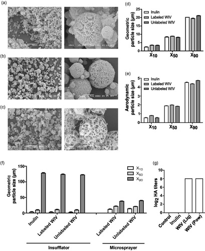
Figure 2. In vivo deposition of labeled liquid and powder WIV formulations. Cotton rats were vaccinated via the pulmonary route with labeled liquid and powder influenza vaccine formulations. Immediately after administration, animals were sacrificed and intact lungs were excised and later lungs were dissected into lung lobes for imaging. (a) Representative IVIS images of the intact (top) and dissected (bottom) lungs showing the deposition of liquid and powder WIV. Due to its proximity with the lungs, the heart (indicated by a yellow circle) was used as a negative control. (b) Percentage of the dose delivered in the trachea and five lung lobes (together) for liquid and powder WIV formulations relative to the total dose deposited in the respiratory tract. (c) Relative dose percentage in trachea and individual lung lobes for liquid and powder WIV formulations. Dose percentages for lung lobes were calculated as: (100-Dose deposition in trachea (%)). Relative dose percentages were determined as: dose % in trachea or lung lobe/Weight % of trachea or lung lobe out of the total lung weight. Data are presented as average ± standard error of the mean with levels of significance represented as *p < .05, **p < .01, ***p < .001.
Figure 3. Systemic immune responses after pulmonary immunization. Cotton rats were vaccinated twice on day 0 and day 21 with liquid or powder formulations of WIV. Control cotton rats received SFD inulin powder via pulmonary route. Three weeks after the second vaccination (on the day of challenge), animals were bled and serum was used for determining systemic immune responses represented by (a) IgG titers (of individual cotton rats), (b) MN titers (of individual cotton rats), and (c) HI titers (sera pooled per experimental group). ELISA titers are represented as log10 titers, while MN and HI titers are represented as log2 titers with significance *p < .05. LoD is represented by dashed line at 2.3 for IgG, 4.32 for MN, and 2.59 for HI titers. Data are presented as average ± standard error of the mean [n = 6 for inulin (Pul-Pow) (•), n = 6 for WIV (i.m.) (▪), n = 5 for WIV (Pul-Liq) (▴), and n = 8 for WIV (Pul-Pow) (▾)].
![Figure 3. Systemic immune responses after pulmonary immunization. Cotton rats were vaccinated twice on day 0 and day 21 with liquid or powder formulations of WIV. Control cotton rats received SFD inulin powder via pulmonary route. Three weeks after the second vaccination (on the day of challenge), animals were bled and serum was used for determining systemic immune responses represented by (a) IgG titers (of individual cotton rats), (b) MN titers (of individual cotton rats), and (c) HI titers (sera pooled per experimental group). ELISA titers are represented as log10 titers, while MN and HI titers are represented as log2 titers with significance *p < .05. LoD is represented by dashed line at 2.3 for IgG, 4.32 for MN, and 2.59 for HI titers. Data are presented as average ± standard error of the mean [n = 6 for inulin (Pul-Pow) (•), n = 6 for WIV (i.m.) (▪), n = 5 for WIV (Pul-Liq) (▴), and n = 8 for WIV (Pul-Pow) (▾)].](/cms/asset/46ff86fa-be34-4c81-ada3-d87a6309f582/idrd_a_1435748_f0003_b.jpg)
Figure 4. Mucosal immune responses after pulmonary immunization. Cotton rats were vaccinated twice followed by a homologous challenge with 107 TCID50/ animal of A/Cal/2009. One day post challenge some animals were sacrificed, nasal, and lung washes were collected. Mucosal immune responses are represented as (a) Nose IgA titers,(b) Lung IgA titers, (c) Lung IgG titers, and (d) MN titers from lung washes pooled per group. Titers are represented as log10 titers along with the significance indicated as *p < .05 and **p < .01. LoD is represented by a dashed line at 0.3 for both nose and lung IgA, while at 1.7 for lung IgG, and at 2 for lung MN titers. Data are presented as average ± standard error of the mean [n = 6 for inulin (Pul-Pow) (•), n = 6 for WIV (i.m.) (▪), n = 5 for WIV (Pul-Liq) (▴), and n = 8 for WIV (Pul-Pow) (▾)].
![Figure 4. Mucosal immune responses after pulmonary immunization. Cotton rats were vaccinated twice followed by a homologous challenge with 107 TCID50/ animal of A/Cal/2009. One day post challenge some animals were sacrificed, nasal, and lung washes were collected. Mucosal immune responses are represented as (a) Nose IgA titers,(b) Lung IgA titers, (c) Lung IgG titers, and (d) MN titers from lung washes pooled per group. Titers are represented as log10 titers along with the significance indicated as *p < .05 and **p < .01. LoD is represented by a dashed line at 0.3 for both nose and lung IgA, while at 1.7 for lung IgG, and at 2 for lung MN titers. Data are presented as average ± standard error of the mean [n = 6 for inulin (Pul-Pow) (•), n = 6 for WIV (i.m.) (▪), n = 5 for WIV (Pul-Liq) (▴), and n = 8 for WIV (Pul-Pow) (▾)].](/cms/asset/dda645cd-f108-4d5a-a864-2e768fd3fe96/idrd_a_1435748_f0004_b.jpg)
Figure 5. Effect of pulmonary vaccination on lung virus titers upon live virus challenge. Three weeks after the second vaccination cotton rats were challenged with 107 TCID50/ animal of A/Cal/2009 virus. One day post challenge some animals were sacrificed and their lungs were homogenized to determine virus load. Virus titers are represented as log10 titers per gram of lung and significant differences between titers of different groups are represented as *p < .05. LoD is represented by a dashed line at 1.3. Data are presented as average ± standard error of the mean [n = 6 for inulin (Pul-Pow) (•), n = 6 for WIV (i.m.) (▪), n = 5 for WIV (Pul-Liq) (▴), and n = 8 for WIV (Pul-Pow) (▾)].
![Figure 5. Effect of pulmonary vaccination on lung virus titers upon live virus challenge. Three weeks after the second vaccination cotton rats were challenged with 107 TCID50/ animal of A/Cal/2009 virus. One day post challenge some animals were sacrificed and their lungs were homogenized to determine virus load. Virus titers are represented as log10 titers per gram of lung and significant differences between titers of different groups are represented as *p < .05. LoD is represented by a dashed line at 1.3. Data are presented as average ± standard error of the mean [n = 6 for inulin (Pul-Pow) (•), n = 6 for WIV (i.m.) (▪), n = 5 for WIV (Pul-Liq) (▴), and n = 8 for WIV (Pul-Pow) (▾)].](/cms/asset/8c88589b-577c-42ff-aff8-d4658f6282e3/idrd_a_1435748_f0005_b.jpg)
Figure 6. Effect of pulmonary vaccination on clinical symptoms after live influenza virus challenge. Upon vaccination and homologous live virus challenge, cotton rats were followed for 10 days for assessment of weight loss (a) Inulin (Pul-Pow), (b) WIV (i.m.), (c) WIV (Pul-Liq), and (d) WIV (Pul-Pow). Also, change in the BF was evaluated for all the animals that were followed (e) Inulin (Pul-Pow), (f) WIV (i.m.), (g) WIV (Pul-Liq), and (h) WIV (Pul-Pow). Day wise BF was plotted for (i) day 1 and (j) day 2 post challenge for animals in each group. Data [(i),( j)] are presented as average ± standard error of the mean . For a to h, each line represents 1 animal. For each figure, •: animal 1; ▪: animal 2; ▴: animal 3. For i and j [n = 3 for inulin (Pul-Pow) (•), n = 3 for WIV (i.m.) (▪), n = 3 for WIV (Pul-Liq) (▴), and n = 3 for WIV (Pul-Pow)(▾)].
![Figure 6. Effect of pulmonary vaccination on clinical symptoms after live influenza virus challenge. Upon vaccination and homologous live virus challenge, cotton rats were followed for 10 days for assessment of weight loss (a) Inulin (Pul-Pow), (b) WIV (i.m.), (c) WIV (Pul-Liq), and (d) WIV (Pul-Pow). Also, change in the BF was evaluated for all the animals that were followed (e) Inulin (Pul-Pow), (f) WIV (i.m.), (g) WIV (Pul-Liq), and (h) WIV (Pul-Pow). Day wise BF was plotted for (i) day 1 and (j) day 2 post challenge for animals in each group. Data [(i),( j)] are presented as average ± standard error of the mean . For a to h, each line represents 1 animal. For each figure, •: animal 1; ▪: animal 2; ▴: animal 3. For i and j [n = 3 for inulin (Pul-Pow) (•), n = 3 for WIV (i.m.) (▪), n = 3 for WIV (Pul-Liq) (▴), and n = 3 for WIV (Pul-Pow)(▾)].](/cms/asset/33ff21b1-3702-433a-8250-c3a0dd18afaf/idrd_a_1435748_f0006_b.jpg)