Figures & data
Table 1. Experimental design of inhaled vs. IV topotecan for the treatment of lung tumors in rats.
Figure 1. Physical properties of the spray-dried powder of topotecan. (A) Scanning Electron Microscope image. (B) Volume particle size distribution (Malvern–Wet Method) and (C) mass particle size distribution recovered from the inhalation chamber.
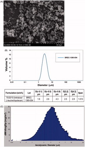
Figure 2. Pharmacokinetic Analysis of Inhaled versus IV topotecan. The levels of topotecan detected in (A) plasma and (B) lung tissue, and (C) the average values from non-compartmental analysis of concentration vs. time are shown.
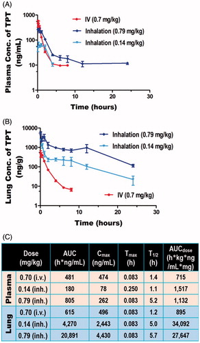
Figure 3. Efficacy of inhaled versus IV topotecan for lung cancer therapy. The survival (A,B) and lung tumor burden (C,D) of rats with the fast-growing H1975 (A and C) and moderately growing A549 (B and D) derived lung cancer were compared between treatment groups.
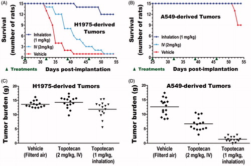
Table 2. Inhaled topotecan is more effective in treating lung cancer than higher IV dose.
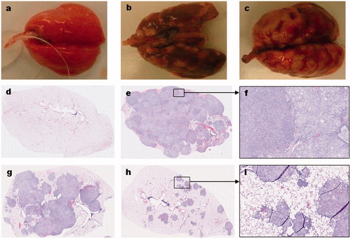
Figure 4. Gross and histological features of orthotopic lung tumors in rats. The gross (a–c) and microscopic (d–i) features of normal lung (a,d), A549-derived tumors (b,e–i), and H1975-derived tumors (c) are shown. Compared to the normal lungs (a) of an age-matched, cancer-free, and treatment naïve rat; the A549 (b) and H1975 (c) derived tumor-bearing lungs from untreated control animals have grossly visible, multinodular, tan-gray, masses that bulge from the pleural and visceral surfaces of the lungs, respectively. Microscopic evaluation of hematoxylin and eosin (H&E) stained sections from A549-derived tumor-bearing lungs (e–i) revealed that the lung parenchyma, smaller airways, and blood vessels are invaded by patches of cancer cells. In a vehicle-treated animal, the entire lung lobe is filled with tumor cells of mainly two types (a smaller or larger cytoplasm) and very little normal airspace is left (e,f). In a rat treated with IV topotecan, about half the lung lob is filled with patches of cancer cells that started to coalesce (g). In contrast, about 90% of the lung parenchyma of a rat treated with inhaled topotecan remained normal and the tumor nodules are much smaller and mostly noncoalescent (h,i).