Figures & data
Figure 1. Schematic diagram of the brain-targeting mixed micellar delivery system (RVG29-Nano-BAP85).
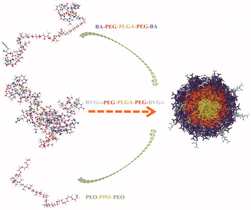
Figure 2. Cellular uptake (A) and antibacterial efficiency (B) of RVG29-Nano-BAP85 with different loading content of RVG29-PEG-PLGA-PEG-RVG29 (mean ± SD, n = 6). Flow cytometry analysis of the RVG29-Nano-BAP85 mixed micelles with different loading content of Pluronic® P85 taken up by BCECs (mean ± SD, n = 6) (C). The concentration of the copolymer mixture was 10 mg/mL.
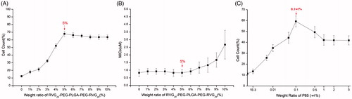
Figure 3. TEM images of RVG29-Nano-BAP85 (A), RVG29-Nano-BA (B), Nano-BAP85 (C), and Nano-BA (D).
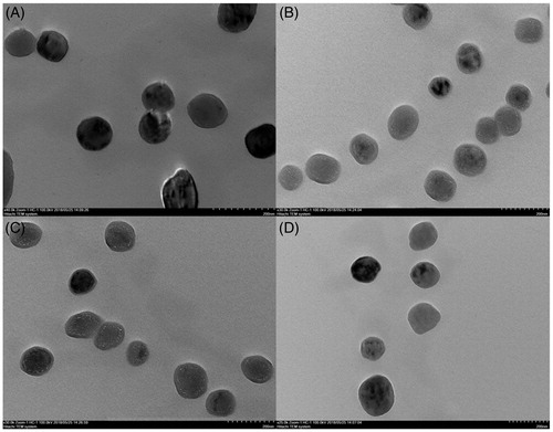
Figure 4. Qualitative (A) and quantitative (B) cellular uptake of FITC-labeled BA, FITC-loaded RVG29-Nano-BAP85, RVG29-Nano-BA, Nano-BAP85, Nano-BA, and PEGylated Nano-BA12K on BCECs for 2 h. The permeability of FITC-labeled BA, FITC-loaded RVG29-Nano-BAP85, RVG29-Nano-BA, Nano-BAP85, Nano-BA, and PEGylated Nano-BA12K across BCECs monolayer (C). The effects of RVG29-Nano-BAP85, RVG29-Nano-BA, Nano-BAP85, Nano-BA, PEGylated Nano-BA12K, and Pluronic® P85 unimers on the intracellular ATP level (D) and microviscosity (E) of BCECs. *** p < .001, significance represents RVG29-Nano-BAP85 vs. RVG29-Nano-BA, Nano-BAP85, Nano-BA, and PEGylated Nano-BA12K. **p < .01; ***p < .001, significance represents RVG29-Nano-BAP85 vs. RVG29-Nano-BA, Nano-BAP85, Nano-BA, and PEGylated Nano-BA12K.
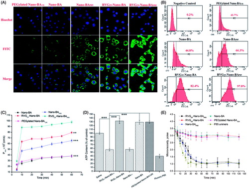
Figure 5. The in vivo noninvasive images of time-dependent whole body imaging of PM mice co-infected with S. pneumonia ATCC 49619 and S. pneumonia 16167 after i.v. injection of DiR-loaded RVG29-Nano-BAP85, RVG29-Nano-BA, Nano-BAP85, Nano-BA, and PEGylated Nano-BA12K (A). The ex vivo optical images of the brain (B) and other organs (C) of the PM mice co-infected with S. pneumonia ATCC 49619 and S. pneumonia 16167 after i.v. injection of DiR-loaded RVG29-Nano-BAP85, RVG29-Nano-BA, Nano-BAP85, Nano-BA, and PEGylated Nano-BA12K. Quantitative analysis for accumulation in the brain of BA in the PM mice co-infected with S. pneumonia ATCC 49619 and S. pneumonia 16167 at different times after intravenous administration of RVG29-Nano-BAP85, RVG29-Nano-BA, Nano-BAP85, Nano-BA, and PEGylated Nano-BA12K at a dose of 30 mg/kg, respectively (D).
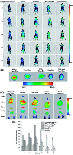
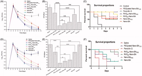
Figure 6. Colony formation unit of S. pneumonia ATCC 49619 in CSF of the mice without any treatment (the negative control group) or treated with Nano-BAs or Penicillin G for various periods of time (A). Quantitative counts of S. pneumonia ATCC 49619 per gram of brain tissues of the mice without any treatment (the control group) or treated with Nano-BAs or Penicillin G for 14 days (B). Survival rate of the PM mice infected with S. pneumonia ATCC49619 after treated with Saline, PEGylated Nano-BA12K, Penicillin G, RVG29-Nano-BAP85, RVG29-Nano-BA, Nano-BAP85, and Nano-BA, respectively (C). Colony formation unit of S. pneumonia 16167 in CSF of the mice without any treatment (the negative control group) or treated with Nano-BAs or Penicillin G for various periods of time (D). Quantitative counts of S. pneumonia 16167 per gram of brain tissues of the mice without any treatment (the control group) or treated with Nano-BAs or Penicillin G for 14 days (E). Survival rate of the PM mice infected with S. pneumonia 16167 after treated with Saline, PEGylated Nano-BA12K, Penicillin G, RVG29-Nano-BAP85, RVG29-Nano-BA, Nano-BAP85, and Nano-BA, respectively (F). Values are mean ± SD of six independent observations. *p < .05, **p < .01, ***p < .001, significance represents RVG29-Nano-BAP85 vs. RVG29-Nano-BA, Nano-BAP85, Nano-BA, and PEGylated Nano-BA12K.