
Figures & data
Figure 1. Temperature sweep. Data are plotted as triplicate samples of SAIB DDS (w/w) as a function of temperature.
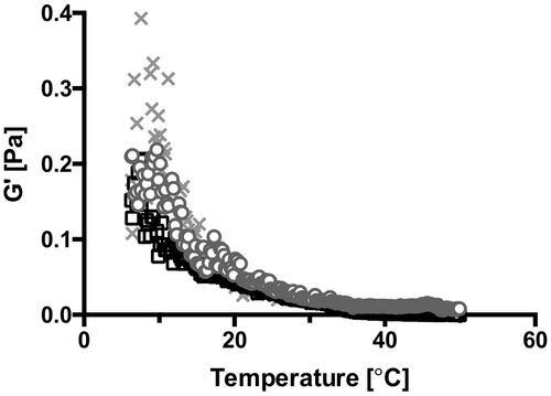
Figure 2. Effect of storage and incubation on the rheological profile of the SAIB DDS (A) and (B) frequency sweep of SAIB DDS stored at different temperatures and measured at either 22 °C (A) or 37 °C (B). (C) G′ after immersion of SAIB DDS in either 10 mM MES buffer or simulated small intestinal fluid (SSIF), both pH 6.5 and measured at 37 °C, and (D) loss on drying after exposure to the same buffer and temperature conditions as described for (C). Data are plotted as mean ± S.D.; n = 3.
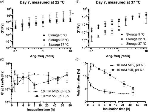
Figure 3. Continuous ramp flow with increasing shear rates from 0 to 25 s–1. Data are plotted as a representative sample chosen from triplicate measurements.
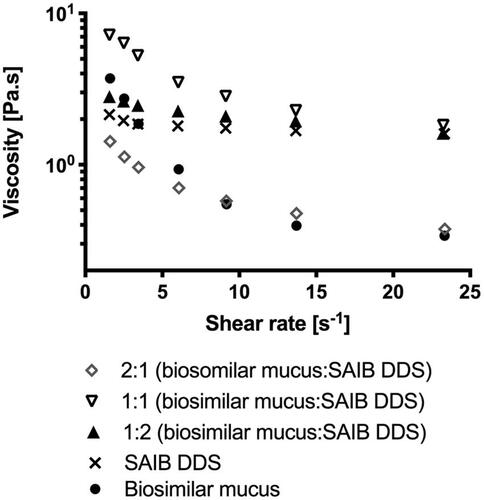
Figure 4. (A) Insulin release from SAIB DDS during the dynamic gastric model experiment simulating fasted state in vivo conditions. The image is obtained using light microscopy with a magnification of 20×. (B) Release of insulin at 37 °C from SAIB DDS after immersion in either 10 mM MES buffer or simulated small intestine fluid (SSIF), both pH 6.5. Data are plotted as mean ± S.D.; n = 3. The images are obtained using light microscopy with a magnification of 100×.
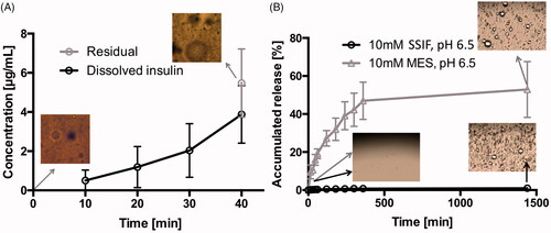
Figure 5. (A) Permeation of 3H-mannitol across a Caco-2 cell monolayer and permeation of 3H-mannitol when incorporated into SAIB DDS. (B) Cell viability (black bar) and epithelial integrity (open bar), assessed by transepithelial electrical resistance (TEER) of Caco-2 cell monolayers after application of SAIB DDS with either 3H-mannitol incorporated into SAIB DDS or co-administered with SAIB DDS for 4 h. Data are plotted as mean ± S.D.; n = 3.
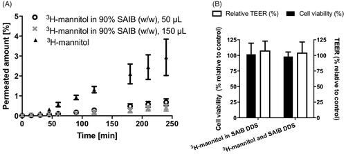
Figure 6. Representative SPECT/CT images showing the biodistribution of orally administered 123I-insulin 10 min, 2 h and 22 h after administration. The scale bar is the same for all images.
