19,632
Views
221
CrossRef citations to date
0
Altmetric
Research Article

PLGA-based biodegradable microspheres in drug delivery: recent advances in research and application

, , , , ORCID Icon, , & ORCID Icon show all
Pages 1397-1418 | Received 16 Apr 2021, Accepted 31 May 2021, Published online: 29 Jun 2021

Figures & data

Table 1. Effects of PLGA composition on particle size, drug loading, and release profile of microspheres.

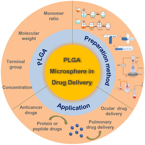
Figure 1. A diagram of PLGA-based biodegradable microspheres in drug delivery.

Figure 1. A diagram of PLGA-based biodegradable microspheres in drug delivery.

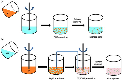
Figure 2. Schematic of PLGA microspheres prepared via (a) single emulsion and (b) double emulsion.

Figure 2. Schematic of PLGA microspheres prepared via (a) single emulsion and (b) double emulsion.

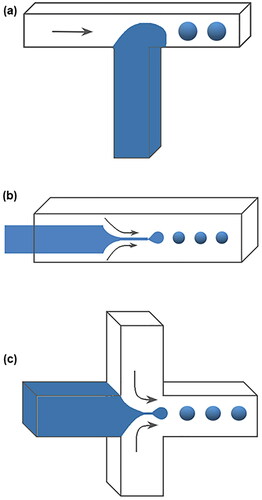
Figure 3. Schematic of microfluidic devices for making PLGA microspheres. (a) T-Junction microfluidic device, (b) co-flow microfluidic device, (c) flow-focusing microfluidic device.

Figure 3. Schematic of microfluidic devices for making PLGA microspheres. (a) T-Junction microfluidic device, (b) co-flow microfluidic device, (c) flow-focusing microfluidic device.

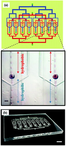
Figure 4. (a) Schematic of a microfluidic parallelization device containing 8 double emulsion drop makers. Hydrophobic channels (indicated in red) and hydrophilic channels (indicated in blue) are located on different layers of the device. Inset shows the simultaneous operation of two adjacent drop makers of the parallel chip. (b) Photograph of the parallelized microfluidic device. Adapted with permission from Reference (Nawar et al., Citation2020). Copyright 2020, Royal Society of Chemistry.

Figure 4. (a) Schematic of a microfluidic parallelization device containing 8 double emulsion drop makers. Hydrophobic channels (indicated in red) and hydrophilic channels (indicated in blue) are located on different layers of the device. Inset shows the simultaneous operation of two adjacent drop makers of the parallel chip. (b) Photograph of the parallelized microfluidic device. Adapted with permission from Reference (Nawar et al., Citation2020). Copyright 2020, Royal Society of Chemistry.

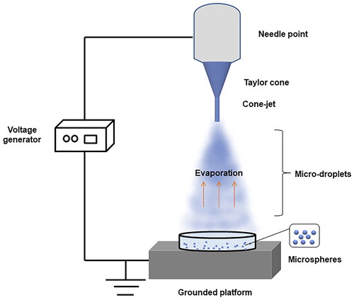
Figure 5. Schematic of the electrospray process for preparing PLGA microspheres.

Figure 5. Schematic of the electrospray process for preparing PLGA microspheres.

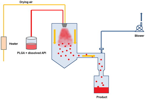
Figure 6. Schematic diagram of the spray drying process.

Figure 6. Schematic diagram of the spray drying process.

Table 2. PLGA-based biodegradable microspheres for cancer drug delivery.

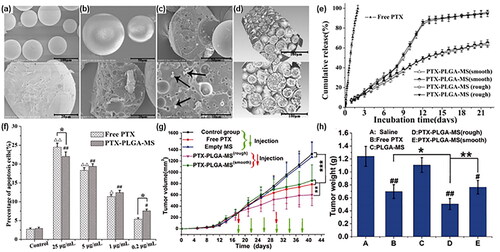
Figure 7. (a) The SEM micrographs of smooth PTX-PLGA-MS. (b-d) The SEM micrographs of rough PTX-PLGA-MS and the arrows represent the PTX drug substances. (e) In-vitro cumulative released curves of free PTX and PTX-PLGA-MS. (f) In-vitro evaluation for apoptosis when PTX formulations co-cultured with U251 cells. (g) Tumor growth curves of smooth or rough PTX-PLGA-MS, PLGA-MS, free PTX, and with no treatment during the entire experiment. (h) Tumor weight of each treatment group at the end of the tests. Adapted with permission from Zhang et al. (Citation2018). Copyright 2018, Informa UK Limited.

Figure 7. (a) The SEM micrographs of smooth PTX-PLGA-MS. (b-d) The SEM micrographs of rough PTX-PLGA-MS and the arrows represent the PTX drug substances. (e) In-vitro cumulative released curves of free PTX and PTX-PLGA-MS. (f) In-vitro evaluation for apoptosis when PTX formulations co-cultured with U251 cells. (g) Tumor growth curves of smooth or rough PTX-PLGA-MS, PLGA-MS, free PTX, and with no treatment during the entire experiment. (h) Tumor weight of each treatment group at the end of the tests. Adapted with permission from Zhang et al. (Citation2018). Copyright 2018, Informa UK Limited.

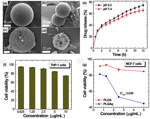
Figure 8. The SEM micrographs of PLGA (a-b) and Quercetin-loaded PLGA (PLGAq) microspheres (c–d). (e) In-vitro cumulative released curves of PLGAq under normal and tumor microenvironment. (f) In-vitro cytotoxicity assay tested using THP-1 cell lines. (g) In-vitro cytotoxicity of PLGA and PLGAq tested on MCF-7 cell lines. Adapted with permission from Karthick et al. (Citation2019). Copyright 2019, Elsevier.

Figure 8. The SEM micrographs of PLGA (a-b) and Quercetin-loaded PLGA (PLGAq) microspheres (c–d). (e) In-vitro cumulative released curves of PLGAq under normal and tumor microenvironment. (f) In-vitro cytotoxicity assay tested using THP-1 cell lines. (g) In-vitro cytotoxicity of PLGA and PLGAq tested on MCF-7 cell lines. Adapted with permission from Karthick et al. (Citation2019). Copyright 2019, Elsevier.

Table 3. PLGA-based biodegradable microspheres for protein or peptide drug delivery.

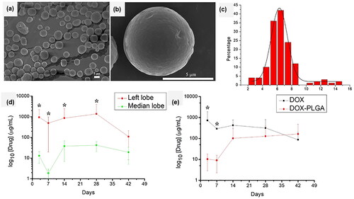
Figure 9. (a, b) The SEM micrographs of microspheres. (c) The size distribution of microspheres. (d) The concentration distribution of DOX in the left and median hepatic lobes of the rats during the experiment. (e) The plasma concentration of DOX in rats after injection of the drug (DOX) – loaded microsphere and free DOX during the experiment. Adapted with permission from Hsu et al. (Citation2020). Copyright 2020, Elsevier.

Figure 9. (a, b) The SEM micrographs of microspheres. (c) The size distribution of microspheres. (d) The concentration distribution of DOX in the left and median hepatic lobes of the rats during the experiment. (e) The plasma concentration of DOX in rats after injection of the drug (DOX) – loaded microsphere and free DOX during the experiment. Adapted with permission from Hsu et al. (Citation2020). Copyright 2020, Elsevier.

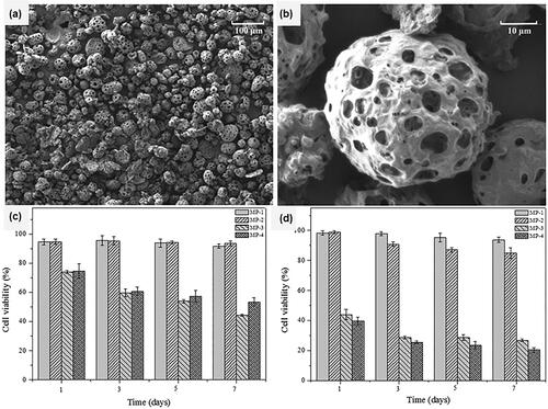
Figure 10. The SEM micrographs of (a) porous PLGA microspheres loaded with doxorubicin and PEI25K/miR-519c (MP-4) and (b) the porous surface, and in-vitro cell viability evaluation of A549 cells treated with the release supernatants from different types of porous PLGA microparticles for 24 h (c) and 48 h (d). Adapted from with permission Wu et al. (Citation2016). Copyright 2016, American Chemical Society.

Figure 10. The SEM micrographs of (a) porous PLGA microspheres loaded with doxorubicin and PEI25K/miR-519c (MP-4) and (b) the porous surface, and in-vitro cell viability evaluation of A549 cells treated with the release supernatants from different types of porous PLGA microparticles for 24 h (c) and 48 h (d). Adapted from with permission Wu et al. (Citation2016). Copyright 2016, American Chemical Society.

Table 4. PLGA-based biodegradable microspheres for pulmonary drug delivery.

Table 5. PLGA-based biodegradable microspheres for ocular drug delivery.