Figures & data
Figure 1. Preparation and characterization of IR780-in-liposome-in-hydrogels (IR780/lipo/gel). (A) Schematic illustration of IR780/lipo/gel. (B) Hydrodynamic size (diameter, nm) and PDI of IR780 liposomes (IR780/lipo). (C) Representative TEM images of IR780/lipo (scale bar: 100 nm). (D) Representative SEM images of IR780/lipo/gel (scale bar: 200 μm). (E) Rheological characterization of IR780/lipo/gel. The storage modulus G′ and loss modulus G″ were plotted logarithmically against frequency (0.1–10 Hz at 37 °C). (F) Absorption spectra of free IR780, IR780/lipo, and IR780/lipo/gel. (G) In vitro release of liposomes from the hydrogel. Data are presented as the means ± SD (n = 3).
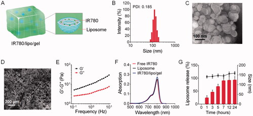
Figure 2. In vivo fluorescence imaging and biodistribution of photosensitizers in CT-26 tumor-bearing mice. (A) In vivo dye fluorescence images in CT-26 tumor-bearing mice at different time after intravenous injection (i.v.) of IR780/lipo and IR792/lipo. In vivo dye fluorescence images in CT-26 tumor-bearing mice after topically application of IR780/lipo/gel and IR792/lipo/gel onto the tumor (B) and the back (C) of mice. (D) F.I. of IR780 and IR792 in the tumors. Data are presented as the means ± SD (n = 3).
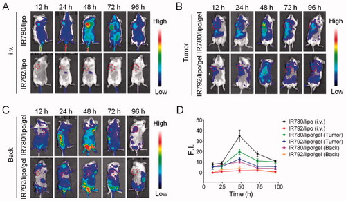
Figure 3. Organ distribution of dye. (A) Representative ex vivo fluorescence images of major organs and tumors after i.v. and topically application of IR780/lipo/gel and IR792/lipo/gel onto the tumor and the back of mice at 72 h. (B) F.I. of IR780 and IR792 in the major organs and tumors. (C) IR792 and IR780 fluorescence imaging in tumor sections following topically application of IR792/lipo/gel and IR780/lipo/gel onto the back. Data are presented as the means ± SD (n = 3).
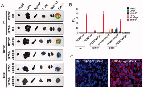
Figure 4. In vivo fluorescence imaging and biodistribution of photosensitizers in experimental lung metastasis model. (A) In vivo dye fluorescence images in experimental lung metastasis model or normal mice at 48 h after i.v. of IR780/lipo, IR792/lipo and topically application of IR780/lipo/gel and IR792/lipo/gel onto the back of mice. (B) Representative ex vivo fluorescence images and F.I. of IR780 and IR792 of major organs and tumors from the mice in (A). Data are presented as the means ± SD (n = 3).
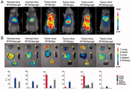
Figure 5. Photothermal anticancer therapy. (A) Antitumor PTT activity of IR780/lipo/gel and IR792/lipo/gel following topically application onto the back of mice in CT-26 colon cancer model. Outliers were removed to improve accuracy of the statistical results. (B) Images of excised tumors on day 14 after treatment. (C) Body weight changes over the treatment period. Data are presented as the means ± SD (n = 6). **p< .01, vs. the indicated groups.
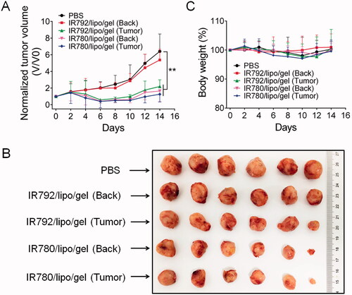
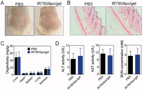
Figure 6. Safety of IR780/lipo/gel. (A) Skin morphology after topically application of IR780/lipo/gel. (B) The skin sections were further examined after H&E staining. (C) Weight ratios of organs to the total body weight and (D) quantification of ALT, AST, and BUN in the plasma after treatment with IR780/lipo/gel.