Figures & data
Figure 1. Illustrating the various inhalational approaches for lung cancer. Reproduced with permission from (Lee et al., Citation2018).
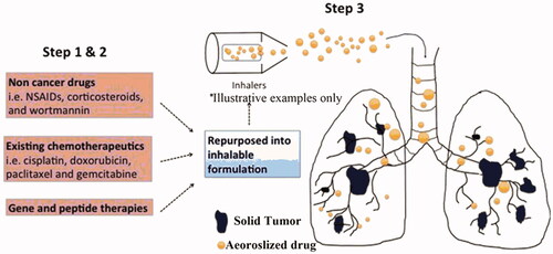
Figure 2. Illustrating the surface-engineered smart nanocarriers and targeted inhalational lung cancer chemotherapy (Anderson et al., Citation2020).
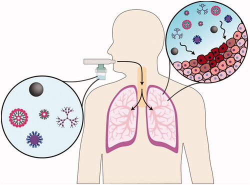
Figure 3. In vivo tumor distribution in the M109 model after inhalation of coated fluorescent folate-grafted copolymer nanocarriers. Confocal images of control untreated M109 mouse lung and coated fluorescent folate-grafted copolymer nanocarriers-treated mouse lung. Green: 25-NBD-cholesterol labeling SLN; red: vessels labeled with isolectinB4; blue: Alexa Fluor 405 labeling the coating. Reproduced with permission from (Rosiere et al., Citation2018). Copyright (2018) American Chemical Society.
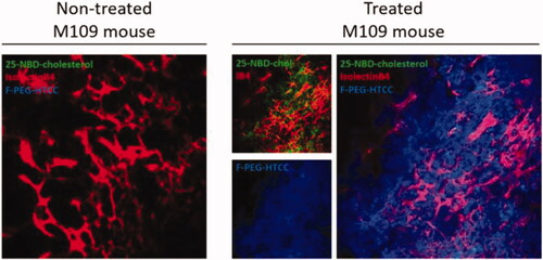
Figure 4. Imaging evaluation of the orthotopic lung cancer model. (A) Bioluminescence optical imaging of control mouse and lung tumor mice of various sizes. (B–D) Magnetic resonance imaging of the control mouse (B) and lung tumor mice of various sizes (C, D). Healthy lung tissues (red) and lung tumors (blue) are displayed (D). (E) Optical imaging of excised organs. (F, G) Computed tomography images of a control mouse (F) and mouse with lung tumors (G). (H) Visualization of lung tumor by the ultrasound imaging system. Reproduced with permission from (Taratula et al., Citation2013).
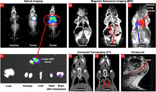
Table 1. Representative preclinical and clinical studies showing the safety and efficacy of inhalational smart nanocarriers in various lung cancers.
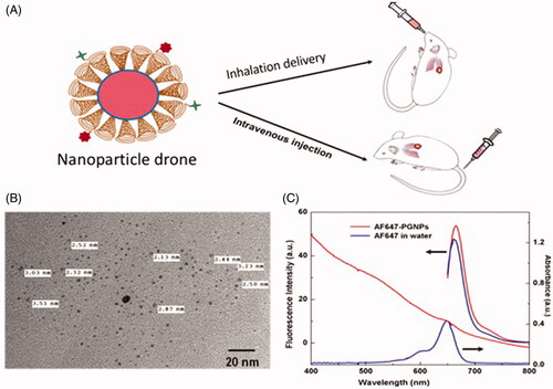
Figure 5. Illustration showing both intravenous and inhalation (INH) delivery of nanoparticle drones; (B) TEM image of lung tumor-targeted with drones; and (C) absorption spectra of drone technology uniquely customized for INH delivery to lung tumors (Ngwa et al., Citation2017).