Figures & data
Figure 1 Micropipette aspiration system measuring the adhesive force of a single endothelial cell.
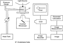
Figure 2 Schematic drawing of the parallel-plate system. The stent coated by endothelial cells was mounted in the parallel plate flow chamber. The hydrostatic pressure head established between the two reservoirs provided a constant volume flow rate through the flow chamber.
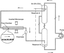
Table 1. Effect of different concentrations of substrates on adhesive forces of HUVECs
Figure 3 Adhesive forces of the HUVEC on the surface of the stents coated with different concentration of the stents coated with different concentrations of PLL and/or FN. A: VEC (Control); B: VEC + 1 µg/ml PLL; C: VEC + 2 µg/ml PLL; D: VEC + 5 µg/mlPLL; E: VEC + 2 µg/ml PLL + 1 µg/ml FN; F: VEC + 2 µg/ml PLL + 2 µg/ml FN; G: VEC + 2 µg/ml PLL + 5 µg/ml FN.
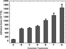
Figure 4 Endothelial cells grown on the surface of the intravascular stents exposed to the perfusion solution.
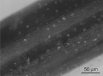
Figure 5 Endothelial cells grown on the surface of the intravascular stents coated by PBS, at the fifth day.
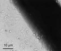
Figure 6 Endothelial cells grown on the surface of the intravascular stents coated by fobronectin (20 mg/ml) at the fifth day.
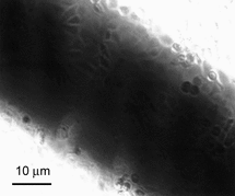
Figure 7 Endothelial cells grown on the surface of the intravascular stents coated by the Poly-l-Lysine at the fifth day.
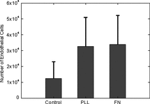
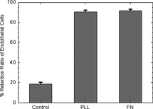
Figure 8 Effect of substrate treatment on the adhesive ratio of endothelial cells under static culture on the stent.
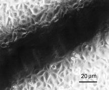
Figure 9 Effect of different substrate treatments on the adhesive ratio of endothelial cells exposed to shear stress on the stent surface.