812
Views
50
CrossRef citations to date
0
Altmetric
Original

Potential Use of Collagen-Chitosan-Hyaluronan Tri-Copolymer Scaffold for Cartilage Tissue Engineering

, , , , &
Pages 27-39 | Published online: 11 Jul 2009

Figures & data

Figure 1 (a) SME micrograph of the porous collagen/chitosan/HA tri-copolymer scaffold. (b) SME micrograph of the porous collagen/hyaluronan/chitosan tri-copolymer scaffold incubation in DMEM medium after 21 days.

Figure 1 (a) SME micrograph of the porous collagen/chitosan/HA tri-copolymer scaffold. (b) SME micrograph of the porous collagen/hyaluronan/chitosan tri-copolymer scaffold incubation in DMEM medium after 21 days.

Table 1. Physico-chemical characteristics of various scaffolds

Figure 2 (a) GAG content of chondrocytes in collagen, collagen/chitosan and collagen/chitosan/HA scaffolds as a function of time of culture. Results are expressed as µg GAG per scaffold. Values are mean ± SD (n = 4); (b) DNA content of chondrocytes in collagen, collagen/chitosan and collagen/chitosan/HA scaffolds as a function of time of culture. Results are expressed as µg DNA per scaffold. Values are mean ± SD (n = 4).
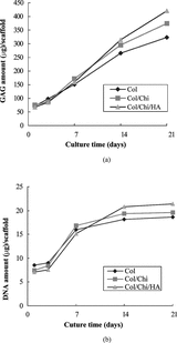
Figure 2 (a) GAG content of chondrocytes in collagen, collagen/chitosan and collagen/chitosan/HA scaffolds as a function of time of culture. Results are expressed as µg GAG per scaffold. Values are mean ± SD (n = 4); (b) DNA content of chondrocytes in collagen, collagen/chitosan and collagen/chitosan/HA scaffolds as a function of time of culture. Results are expressed as µg DNA per scaffold. Values are mean ± SD (n = 4).

Figure 3 (a) Chondrocytes in Col/Chi/HA matrix 1 week after seeding. (b) chondrocytes in Col/Chi/HA matrix 3 weeks after seeding. H&E staining.

Figure 3 (a) Chondrocytes in Col/Chi/HA matrix 1 week after seeding. (b) chondrocytes in Col/Chi/HA matrix 3 weeks after seeding. H&E staining.

Figure 4 (a and b) Environment scanning electron micrographs in Col/Chi/HA scaffold, a week after cell seeding, and at two magnifications. Bar indicates 50 µm in a and 20 µm in b. (c and d) Environment scanning electron micrographs in Col/Chi/HA scaffold 3 weeks after seeding with chondrocytes. (d) Magnification from the same area as C.
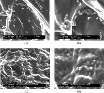
Figure 4 (a and b) Environment scanning electron micrographs in Col/Chi/HA scaffold, a week after cell seeding, and at two magnifications. Bar indicates 50 µm in a and 20 µm in b. (c and d) Environment scanning electron micrographs in Col/Chi/HA scaffold 3 weeks after seeding with chondrocytes. (d) Magnification from the same area as C.

Figure 5 Reverse transcriptase-polymerase chain reaction results against collagen types I and II of chondrocytes according to culture after 14 days within Col/Chi/HA scaffold. 1 = collagen type II and GAPDH; 2 = collagen I and GAPDH; M = 100 bp, 250 bp, 500 bp, 750 bp, 1000 bp and 2000 bp DNA ladder.
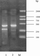
Figure 5 Reverse transcriptase-polymerase chain reaction results against collagen types I and II of chondrocytes according to culture after 14 days within Col/Chi/HA scaffold. 1 = collagen type II and GAPDH; 2 = collagen I and GAPDH; M = 100 bp, 250 bp, 500 bp, 750 bp, 1000 bp and 2000 bp DNA ladder.

Reprints and Corporate Permissions

Please note: Selecting permissions does not provide access to the full text of the article, please see our help page How do I view content?

To request a reprint or corporate permissions for this article, please click on the relevant link below:

Academic Permissions

Please note: Selecting permissions does not provide access to the full text of the article, please see our help page How do I view content?

Obtain permissions instantly via Rightslink by clicking on the button below:

If you are unable to obtain permissions via Rightslink, please complete and submit this Permissions form. For more information, please visit our Permissions help page.