404
Views
7
CrossRef citations to date
0
Altmetric
Original

The Biocompatibility Research of Functional Schwann Cells Induced from Bone Mesenchymal Cells with Chitosan Conduit Membrane

, , , , &
Pages 91-99 | Published online: 11 Jul 2009

Figures & data

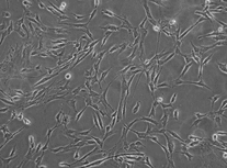
Figure 1 The morphological difference of un-induced MSCs (A) and induced MSCs (B) under phase–contrast microscopy (100×).

Figure 1 The morphological difference of un-induced MSCs (A) and induced MSCs (B) under phase–contrast microscopy (100×).

Figure 2 Immunofluorescence localization of S-100 (A) and GFAP (B) in differentiated MSCs, and the corresponding nuclei counterstained with DAPI (C and D). The red region in cell body was the positive reaction region (100×).
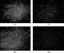
Figure 2 Immunofluorescence localization of S-100 (A) and GFAP (B) in differentiated MSCs, and the corresponding nuclei counterstained with DAPI (C and D). The red region in cell body was the positive reaction region (100×).

Figure 3 Induced MSCs adhered to the chitosan membrane (100×).

Figure 3 Induced MSCs adhered to the chitosan membrane (100×).

Table 1. Absorption value of three groups (x ± s)
Reprints and Corporate Permissions

Please note: Selecting permissions does not provide access to the full text of the article, please see our help page How do I view content?

To request a reprint or corporate permissions for this article, please click on the relevant link below:

Academic Permissions

Please note: Selecting permissions does not provide access to the full text of the article, please see our help page How do I view content?

Obtain permissions instantly via Rightslink by clicking on the button below:

If you are unable to obtain permissions via Rightslink, please complete and submit this Permissions form. For more information, please visit our Permissions help page.