Figures & data
Figure 1 Elution profiles of 3 hours crosslinked PolyHb were obtained by running on a Sephacryl-300 HR 1.6 cm × 70 cm column, equilibrated with 0.1 M Tris · HCl pH 7.5, and eluted at 15 ml/hr. The percentage of molecular weight less than 100 KDa is 78%.
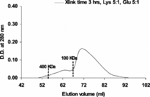
Figure 2 Elution profiles of 24 hours crosslinked PolyHb were obtained by running on a Sephacryl-300 HR 1.6 cm × 70 cm column, equilibrated with 0.1 M Tris · HCl pH 7.5, and eluted at 15 ml/hr. The percentage of molecular weight less than 100 KDa is 38%.
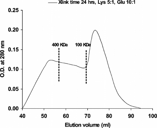
Figure 3 Elution profiles of 24 hours crosslinked PolyHb were obtained by running on a Sephacryl-300 HR 1.6 cm × 70 cm column, equilibrated with 0.1 M Tris · HCl pH 7.5, and eluted at 15 ml/hr. The percentage of molecular weight less than 100 KDa is 16%.
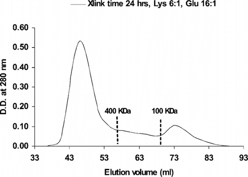
Figure 4 Oxygen dissociation curve of hemoglobin in the free form and PolyHb form. Hemoglobin: free form; 78% LMW PolyHb: contains 78% of low molecular weight molecules (<100 KDa); 38% LMW PolyHb: contains 38% of low molecular weight molecules (<100 KDa); 16% LMW PolyHb: contains 16% of low molecular weight molecules (<100 KDa).
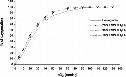
Figure 5 Mean arterial pressure (MAP) with injections of PolyHb samples containing 16%, 38% and 78% of low molecular weight (LMW) components respectively. Injections were started at 30 minutes. All values are represented as mean ±SEM. Preliminary study using PolyHb with 0.4% tetrameric haemoglobin did not show any changes in MAP.
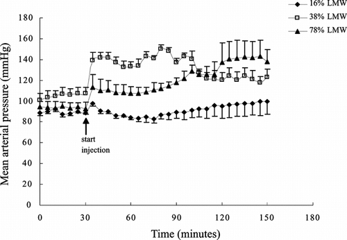
Figure 6 Changes in electrocardiogram (ECG) after injection of 1/6 toploading with PolyHb containing 16%, 38% and 78% of tetramers at 30, 60, 90, and 120 minutes. Pre-IV: before intravenous injection; Post-IV: after intravenous injection. Preliminary study shows that similar toploading using PolyHb with 0.4% tetramers did not result in any changes.
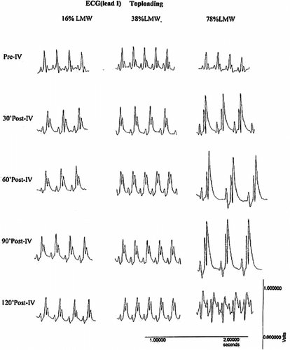
Table 1 Analysis of the total amounts of tetrameric hemoglobin
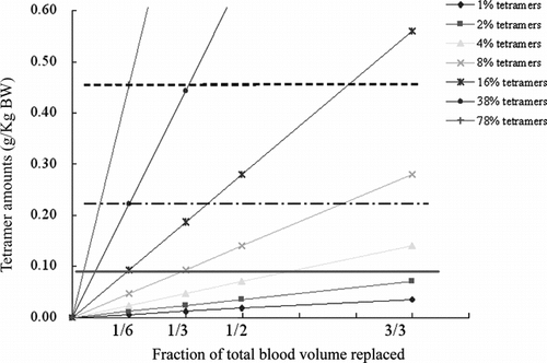
Figure 7 The amount of tetrameric Hb (g/Kg body weight) in PolyHb with 1%, 2%, 4%, 8%, 16%, 38%, and 78% of tetrameric Hb for 1/6, 1/3, 1/2, and 3/3 blood volume replacement. The top dash line represents the tetramer amount (0.455 g/Kg body weight) where severe changes occur in ECG. The middle line shows the tetramer level where blood pressure started to markedly increase (0.222 g/Kg body weight), and this is also the highest concentration that do not cause marked changes in ECG, except for a slight elevation of the ST segment. The bottom horizontal line shows the level of tetrameric Hb (0.093 g/Kg body weight) that did not cause an increase in arterial blood pressure.