Figures & data
Figure 1 An unimplanted AneuRx device fully deployed and manufactured by Medtronic AVE: it consists of a bifurcation with limbs of different lengths, one long and one short, in which an ipsilateral limb is inserted. The thin wall of the polyester is externally supported by self-expanding stents made of shape memory alloy (Nitinol) and sewn by means of multifilament polyester sutures.
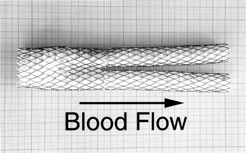
Figure 2 CT scan illustrating the infra-renal abdominal aorta aneurysm of the 75 y.o. patient prior to the stent-graft deployment. The diameter of the aneurysm was 5.7 cm.
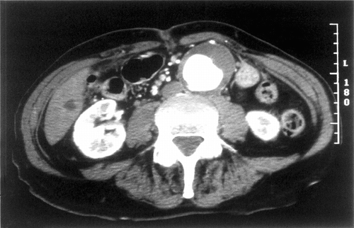
Figure 3 CT scan of the aneurysm of the abdominal aorta fitted with the AneuRx endovascular prosthesis at 18 months. The diameter of the aneurysm decreased in size from 5.7 cm pre-op to 4.6 cm.
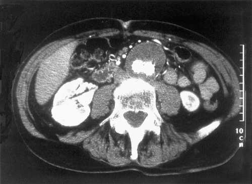
Figure 4 Cross-section of the device together with the reorganized aneurysmal sac where both limbs of the device are still very close, i.e., in the downstream vicinity of the crotch. The lumens of both limbs are clearly delineated (A and A′), whereas the content of the aneurysmal sac has solidified except in a small section (B).
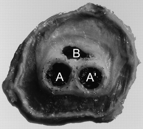
Figure 5 Histology of the cross section slightly upstream of . The stents of the bifurcation of the short limb of the device and the one of the ipsilateral limb are superposed, resulting in the compression of the polyester tube, which loses its cylindrical characteristics (A). On the opposite, the polyester tube preserves its cylindrical shape (A′). The content of the aneurysmal sac has solidified; however, a small canal is still visible (B). The fibrous tissues are almost acellular and the wall of the artery is stretched but mostly keeps its continuity. There are, however, some disruptions.
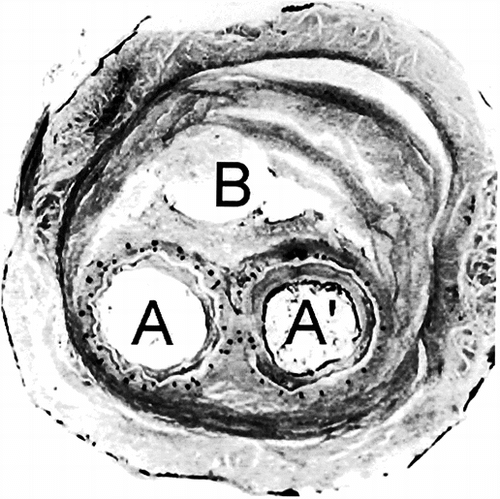
Figure 6 Proton density weighted image of the cross-section of the explanted device with two lumens, A, and A′, corresponding to both limbs of the device. The content of the aneurysmal sac has solidified whereas there is still an open canal likely to let fluids or blood flow.
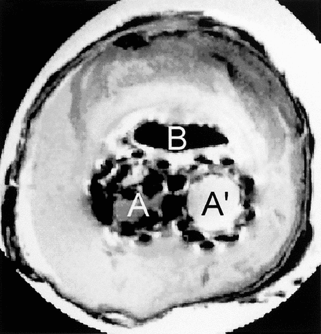
Figure 7 T1 weighted MR image.
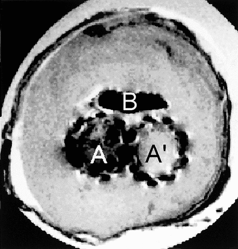
Figure 8 T2 weighted MR images.
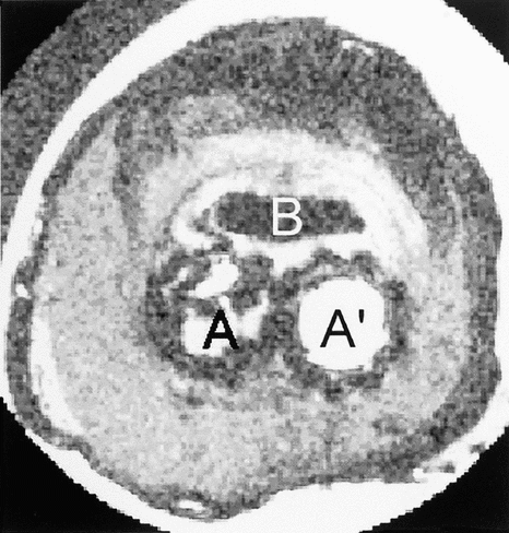
Figure 9 Parametric MR image giving the value of apparent spin-spin relaxations rate R2 in each voxel. The image is windowed between 20 and 100 s−1.
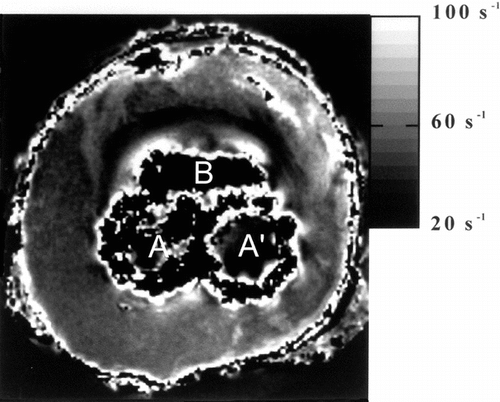
Figure 10 Parametric MR image giving the value of mean diffusivity in each voxel. The image is windowed between 0 and 1.5 × 10⁻³ mm.s−1.
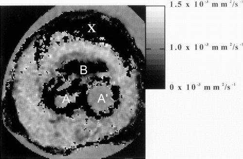
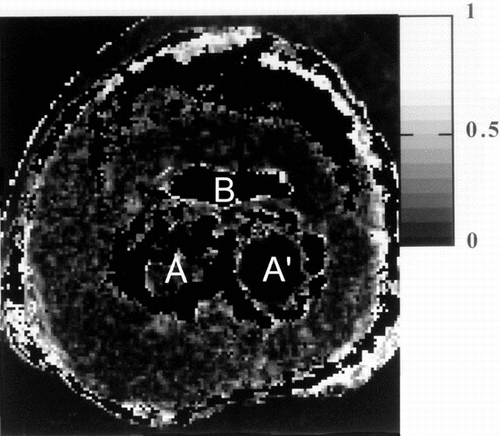
Figure 11 Parametric MR image giving the value of the fractional anisotropy index in each voxel. The image is windowed between 0 and 1.0 corresponding to isotropic diffusion and 1 to infinite anisotropy.