Figures & data
Table 1 The water uptake and size distribution of chitosan beads
Figure 1 Scanning electron micrographs demonstrating the surface morphology of (A) empty and (B) BSA-loaded chitosan-TPP beads. C and D are the surface details of B.
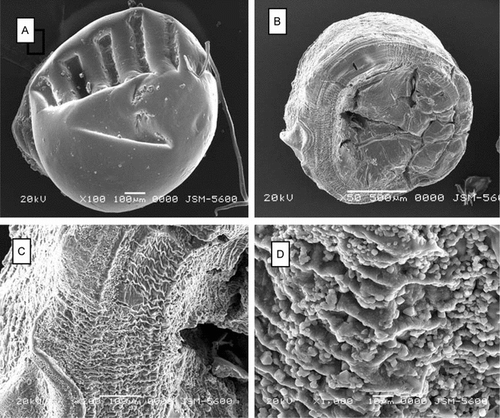
Figure 2 In vitro degradation of chitosan beads in dissolution solutions of PBS at (A) pH 7.4 and (B) pH 6.5 (37°C; static conditions, weekly fresh medium changes). C: chitosan-TPP; CA: alginate-coated chitosan-TPP; CGA: glutaraldehyde-crosslinked chitosan-TPP. All values presented are given as mean ± SD.
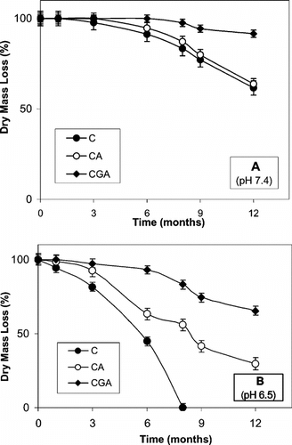
Figure 3 Histological appearance of chitosan beads retrieved at 2 months post-implantation from the epigastric groin fascia of Wistar rats (H&E staining). A and B: chitosan-TPP (C) beads; C and D: alginate-coated chitosan-TPP (CA) beads; E and F: glutaraldehyde-crosslinked chitosan-TPP (CGA) beads. F is the detail of E. FP: fat pad; arrows: capillaries. Scale bars: 200 µm.
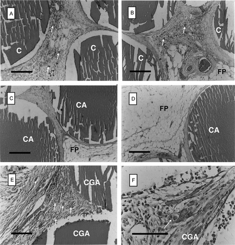
Figure 4 Histological appearance of chitosan beads retrieved at 6 months post-implantation from the epigastric groin fascia of Wistar rats (H&E staining). A and B: chitosan-TPP (C) beads; C and D: alginate-coated chitosan-TPP (CA) beads; E and F: glutaraldehyde-crosslinked chitosan-TPP (CGA) beads. F is the detail of E. FP: fat pad; arrows: capillaries. Scale bars: 200 µm.
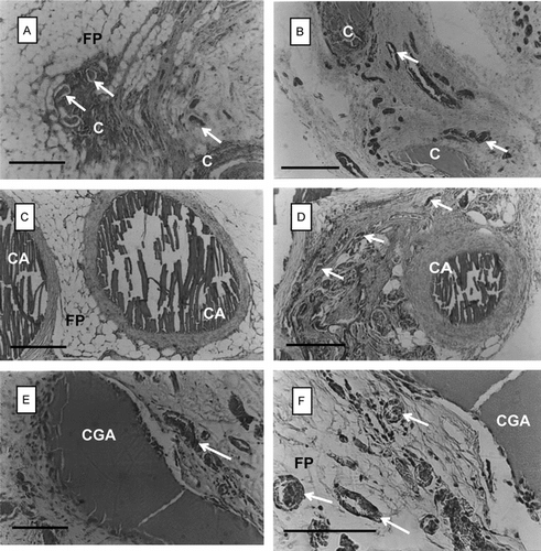
Table 2 Histomorphometric analysis on the in vivo biodegradation of chitosan beadsFootnote[1]