Figures & data
Figure 1 Chemical structure of a single unit of unfractionated heparin (from [Citation[3]]).
![Figure 1 Chemical structure of a single unit of unfractionated heparin (from [Citation[3]]).](/cms/asset/290e4a86-4914-4aa3-814f-3d6b34748c27/ianb19_a_176937_f0001_b.gif)
Figure 2 Schematic diagram of the standard curve obtained from [Citation[7]].
![Figure 2 Schematic diagram of the standard curve obtained from [Citation[7]].](/cms/asset/e72e4b0e-882f-4565-9cea-b50574be1a41/ianb19_a_176937_f0002_b.gif)
Figure 3 Droplet generator.
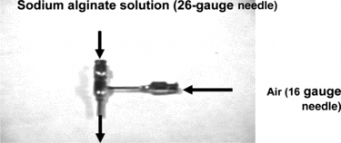
Figure 4 Graph of heparin concentrations during the rate experiment using 8000 beads and 40 ml of 3 U/mL heparin solution.
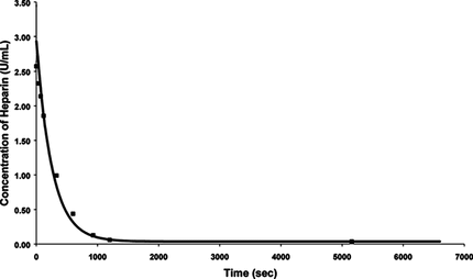
Figure 5 Repeat experiments for 850 beads of 900 µm diameter and 23 mL of 3 U/mL of heparin solution.
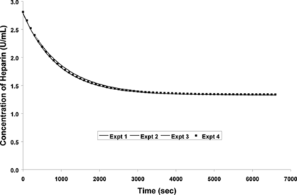
Figure 6 Concentration of heparin in saline solution containing no beads.
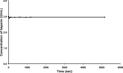
Figure 7 Experimental data fitted to a first order adsorption process.
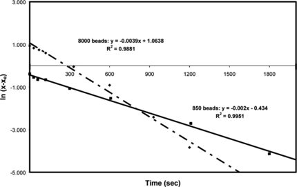
Figure 8 Comparison of rate experiments for different number of beads at 25°C.
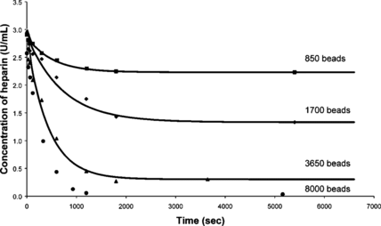
Figure 9 Correlation of K-values to the number of beads.
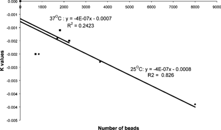
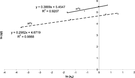
Figure 10 Data points obtained from the rate experiments fitted to Freundlich Isotherm.