Figures & data
Figure 1 Changes in body weight of the Wistar rats during one year following intravenous bolus infusion of HbV at a dose rate of 20 ml/kg. All the rats were housed individually in cages and provided with food and water ad libitum.
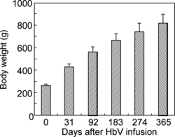
Figure 2 Changes in plasma biochemical parameters after intravenous bolus infusion of HbV into Wistar rats at a dose rate of 20 ml/kg. Data for 7 days (white bars) are cited from our previous report [Citation[8]]. B, baseline.
![Figure 2 Changes in plasma biochemical parameters after intravenous bolus infusion of HbV into Wistar rats at a dose rate of 20 ml/kg. Data for 7 days (white bars) are cited from our previous report [Citation[8]]. B, baseline.](/cms/asset/49ab8fff-00d2-4765-b561-4cd6f27ce509/ianb19_a_197359_f0002_b.gif)
Figure 3 Changes in plasma levels of lipid components, iron, and bilirubin after intravenous bolus infusion of HbV into Wistar rats at a dose rate of 20 ml/kg. Data for 7 days (white bars) are cited from our previous report [Citation[8]]. B, baseline. The values of black bars are from one-year-old rats housed in pairs in cages, not individually (cited from the breeder's data) [Citation[17]].
![Figure 3 Changes in plasma levels of lipid components, iron, and bilirubin after intravenous bolus infusion of HbV into Wistar rats at a dose rate of 20 ml/kg. Data for 7 days (white bars) are cited from our previous report [Citation[8]]. B, baseline. The values of black bars are from one-year-old rats housed in pairs in cages, not individually (cited from the breeder's data) [Citation[17]].](/cms/asset/935681c9-c33f-4804-8ed2-3ad7768a8ed5/ianb19_a_197359_f0003_b.gif)
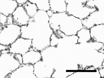
Figure 4 Histology of lung one year after bolus infusion of HbV stained with hematoxylin/eosin. Scale bar, 100 µm.