Figures & data
Figure 1 Effects of HbV infusion on peripheral blood cells. HbV, EV or saline was infused into rats at top-load from the tail vein. Blood was sampled and numbers of red cells, white cells and platelets were counted with an automatic cell counter. Triangles, HbV; squares, EV; circles, saline. N = 3–4, mean±SD.
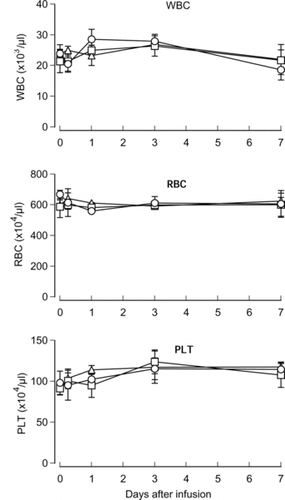
Figure 2 Effects of HbV infusion on the leukocyte population. HbV, EV or saline was infused into rats at top-load from the tail vein. Blood was sampled and ratios of lymphocytes, monocytes and granulocytes were analyzed with the flow cytometer. Triangles, HbV; squares, EV; circles, saline. N = 3–4, mean±SD. *p < 0.05.
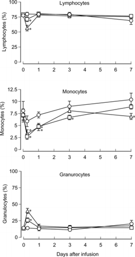
Figure 3 Effects of HbV infusion on the T cell subset. HbV, EV or saline was infused into rats at top-load from the tail vein. Blood was sampled and ratios of CD4+ or CD8+ T cells were analyzed as described in the Materials and Methods section. Black bars, HbV; gray bars, EV; white bars, saline. N = 3–4, mean±SD. *p < 0.05.
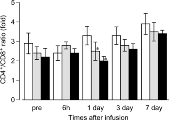
Figure 4 Changes of the complement titer in the rat serum after HbV infusion. HbV, EV or saline was infused into the rats at top-load from the tail vein. Blood was sampled, and sera were prepared as described in the Materials and Methods section. The complement titer was measured and indicated as CH50. Triangles, HbV; squares, EV; circles, saline. N = 3–4, mean ±SD. *p < 0.05.
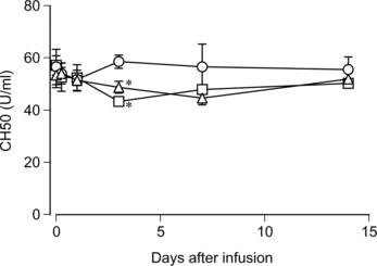
Figure 5 Effects of HbV on the rat complement in vitro. HbV, EV or saline was mixed with rat sera and incubated at 37°C for 1 hr. After centrifugation, the complement titer in the supernatant was measured. Black bars, HbV; gray bars, EV; white bars, saline. N = 5, mean±SD.
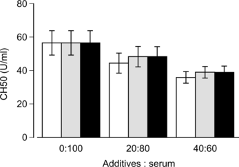
Figure 6 Effects of repeated infusion of HbV on the rat serum complement. HbV or saline was infused into rats at top-load from the tail vein at the time point as indicated by arrows. Blood sampling, sera preparation and complement measurement were carried out as described in the Materials and Methods section. Closed circles, HbV; open circles, saline. N = 3–5, mean±SD. *p < 0.05.
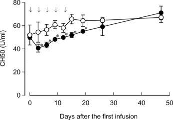
Table 1. Lack of anaphylactic shock in rats infused with EV