Figures & data
Table 1. Experimental groups
Figure 1 Mean arterial pressure changes of rabbits in groups A, B, C, D, E, and F during experiments. MAP decreased after blood withdrawl and increased to normal level when rabbits transfused by autologous whole blood (group A), PEG-bHb containing different concentration of MetHb (groups B, C, D, and E) and dextran (group F), and MAP of rabbits maintained at normal levels until the acute experiment finish except group F.
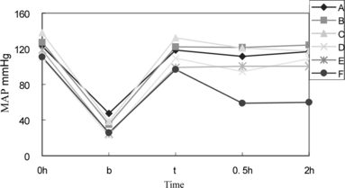
Figure 2 Heart rate changes with hemorrhage transfusion among six group rabbits. Heart rate decreased after rabbits were withdrew 50% volume blood, and recruited when whole blood (group A), PEG-bHb (5% MetHb in group B, 8% MetHb in group C, 15% MetHb in group D, and 25% MetHb in group E) and dextran (group F) were transfused.
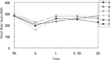
Figure 3 Shown is plasma concentration-time curve of PEG-bHb obtained after rabbits were transfused with PEG-bHb containing 5% MetHb (group B), 8% MetHb (group C), 15% MetHb (group D), and 25% MetHb (group E). Half time of PEG-bHb in four groups was similar.
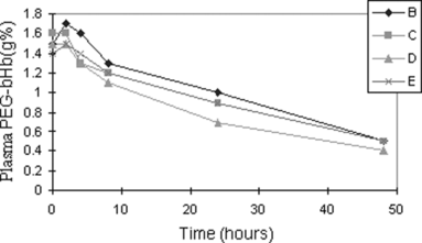
Figure 4 Plasma MetHb concentration changes of rabbits in groups B, C, D, and E transfused by PEG-bHb containing MetHb of 5%, 8%, 15%, and 25%. MetHb concentration decreased quickly during 4 hours after PEG-bHb infusion.
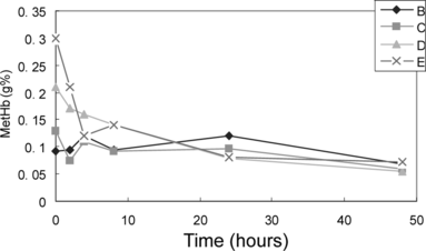
Figure 5 Whole blood Hb concentrations of group B, C, D, E and F rabbits decreased significantly after exchanged transfusion and recovered to about 80% of baseline values at the 8th day.
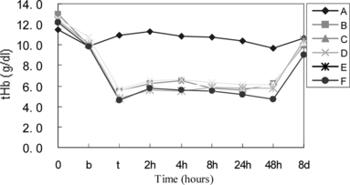
Table 2. Reticular red blood cell counts of six group rabbits during experiments
Figure 6 a) Arterial pH decreased after exchange transfusion and was upregulated to about normal level since 2 hours after fluids of whole blood and PEG-bHb containing different MetHb. Transfusion of dextran did not correct low pH of experiment rabbits quickly. b) Arterial PCO2 decreased during hemorrhage and then changed at the contrary direction as arterial pH. c) Shown is aterial [HCO3–] changes of six group rabbits. It implied dextran transfusion could not improve rabbits ischemia status that low HCO3-level in group F.
![Figure 6 a) Arterial pH decreased after exchange transfusion and was upregulated to about normal level since 2 hours after fluids of whole blood and PEG-bHb containing different MetHb. Transfusion of dextran did not correct low pH of experiment rabbits quickly. b) Arterial PCO2 decreased during hemorrhage and then changed at the contrary direction as arterial pH. c) Shown is aterial [HCO3–] changes of six group rabbits. It implied dextran transfusion could not improve rabbits ischemia status that low HCO3-level in group F.](/cms/asset/551885e9-f88e-4e39-87a6-128e2b2a16ea/ianb19_a_237744_f0006_b.gif)
Figure 7 Blood biochemical markers of CK, ALT, and BUN were used to evaluate the heart, liver, and kidney damage. a): circulation CK increased in rabbits after exchange transfusion, peaked at different time owning to different resuscitation fluids infusion and restored to normal level until 8 d. b): ALT increased in six group rabbits after exchange transfusion and returned to normal at 8 d. c): There is no significant increase in circulating blood urea concentration in groups that exchange transfused by autologous whole blood and PEG-bHb (containing MetHb of 5%, 8%, 15%, and 25%) and recovered at 8 d.
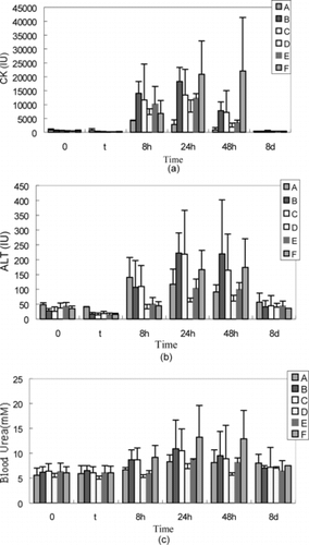
Figure 8 a) Moderate hydropic change and eosinophil granules were observed in group E rabbit liver (× 100). b) moderate vacuolar degeneration was in tubule epthelia cytoplasma of group E rabbits transfused by PEG-bHb containing 25% MetHb (× 100).
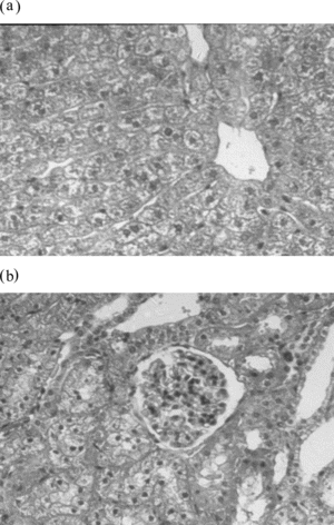
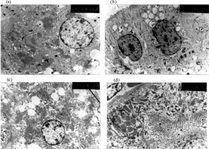
Figure 9 a) Ultrastructure was basically normal except that endoplasmic reticulum dilatation, fat denaturalization and high electron density granules were observed in hepatocytes of rabbits exchange transfused by PEG-bHb containing MetHb of 5%, 8% and 15% (group B, C, and D) (× 4000). b) There're endoplasmic reticulum dilatation and increase of lysosome in renal tululae epithelial cells of group B, C, and D rabbits, other organelles were normal (× 4000). c) Except changes mentioned in group B, C, and D, partial cell organelle collapse occurred in hepatocytes of group E (× 4000). d) Partial cell organelle collapse were also observed in renal tubule epithelial cells of group E rabbits (× 4000).
